. 2015 Mar 26;10(3):e0122297.
doi: 10.1371/journal.pone.0122297. eCollection 2015.

SIRT3 and SIRT5 regulate the enzyme activity and cardiolipin binding of very long-chain acyl-CoA dehydrogenase

Affiliations

SIRT3 and SIRT5 regulate the enzyme activity and cardiolipin binding of very long-chain acyl-CoA dehydrogenase

Yuxun Zhang et al. PLoS One. .

Abstract

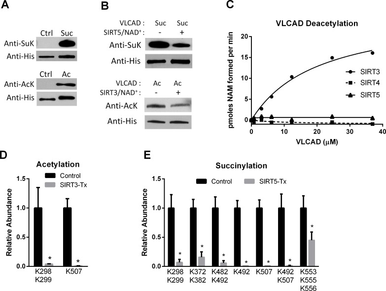
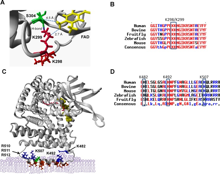
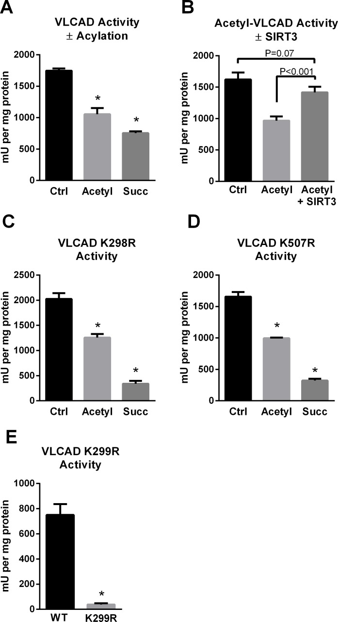
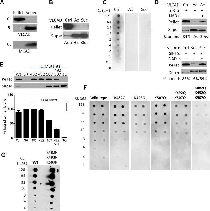
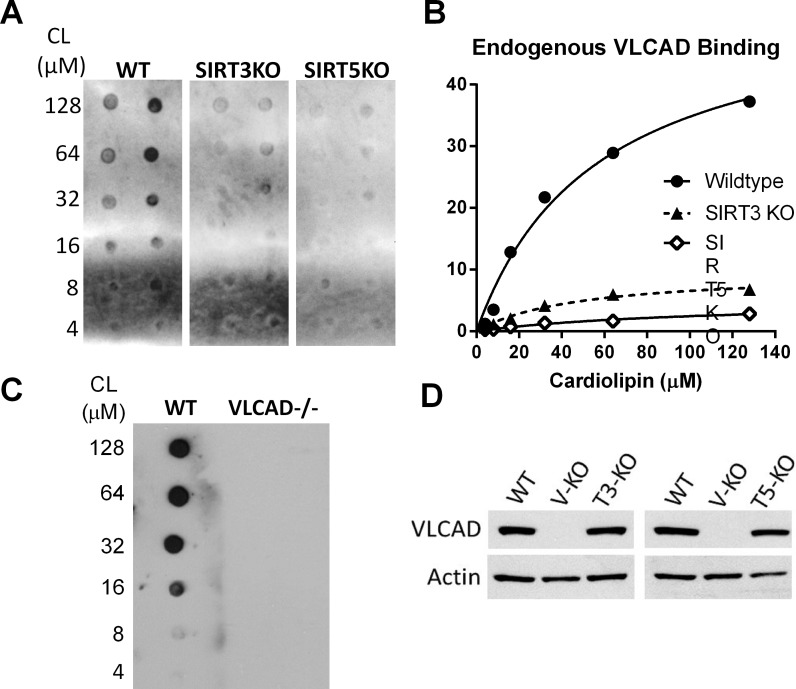
SIRT3 and SIRT5 have been shown to regulate mitochondrial fatty acid oxidation but the molecular mechanisms behind the regulation are lacking. Here, we demonstrate that SIRT3 and SIRT5 both target human very long-chain acyl-CoA dehydrogenase (VLCAD), a key fatty acid oxidation enzyme. SIRT3 deacetylates and SIRT5 desuccinylates K299 which serves to stabilize the essential FAD cofactor in the active site. Further, we show that VLCAD binds strongly to cardiolipin and isolated mitochondrial membranes via a domain near the C-terminus containing lysines K482, K492, and K507. Acetylation or succinylation of these residues eliminates binding of VLCAD to cardiolipin. SIRT3 deacetylates K507 while SIRT5 desuccinylates K482, K492, and K507. Sirtuin deacylation of recombinant VLCAD rescues membrane binding. Endogenous VLCAD from SIRT3 and SIRT5 knockout mouse liver shows reduced binding to cardiolipin. Thus, SIRT3 and SIRT5 promote fatty acid oxidation by converging upon VLCAD to promote its activity and membrane localization. Regulation of cardiolipin binding by reversible lysine acylation is a novel mechanism that is predicted to extrapolate to other metabolic proteins that localize to the inner mitochondrial membrane.


Conflict of interest statement

Competing Interests: The authors have declared that no competing interests exist.

Figures

Fig 1
Fig 1. SIRT3 and SIRT5 deacylate VLCAD at overlapping sites.
A) Recombinant, unmodified VLCAD (Ctrl) was subjected to chemical succinylation (top) or acetylation (bottom) which was verified by western blotting with anti-succinyllysine (SuK) or anti-acetyllysine (AcK) antibodies. B) Chemically succinylated (Suc) and acetylated (Ac) VLCAD proteins were reacted with SIRT5 and SIRT3, respectively. Changes in succinylation or acetylation were then evaluated by western blotting, with anti-His blotting as loading control. C) Only SIRT3 reacts with chemically acetylated VLCAD as determined by incubating increasing amounts of acetylated VLCAD with SIRT3, SIRT4, or SIRT5 in the presence of radiolabeled NAD+. Shown are the means of duplicate assays. D) Acetylated VLCAD was treated with SIRT3 or inactive mutant SIRT3 (Control). Quantitative mass spectrometry was used to determine the relative abundance of acetylated peptides. Shown are acetylation sites with >2-fold change. See S1 Dataset for details. E) Succinylated VLCAD was treated with SIRT5 or inactive mutant SIRT5 (Control) and succinylated peptides were quantified by mass spectrometry. Shown are succinylation sites with >2-fold change. See S2 Dataset for details. D and E both depict the means and standard deviations of quadruplicate assays.
Fig 2
Fig 2. SIRT3 and SIRT5 deacylate lysines that localize to the active site and putative membrane binding domain of VLCAD.
K299 (red) hydrogen bonds with neighboring S304 (green), and both are within interacting distance of the essential FAD cofactor (yellow) which is non-covalently bound in the VLCAD active site. B) Amino acid alignment of the region surrounding K299, showing conservation of this residue across diverse species. C) The portion of VLCAD spanning residues 486–518, which includes sirtuin target sites K492 and K507, is disordered in the crystal structure. PsiPred was used to generate a model of the disordered segment which was overlaid upon the structure of a VLCAD monomer. Hydrophobic residues are rendered red, positively charged residues blue, and negatively charged residues green. The active site is indicated as FAD in yellow and acyl-CoA substrate in red. D) Amino acid alignment of the putative membrane-binding amphipathic helix.
Fig 3
Fig 3. The SIRT3/SIRT5 target site K299 is critical for FAD binding and VLCAD activity.
A) Chemical acetylation and succinylation both reduce enzymatic activity of recombinant VLCAD. B) Incubation of acetylated VLCAD with SIRT3 rescues activity, while incubation of succinylated VLCAD with SIRT5 does not (not shown). C) Mutant K298R retains sensitivity to acylation-induced loss of activity,suggesting that K298 does not play a mechanistic role in the reduced activity. D) Likewise, mutant K507R retains sensitivity to acylation-induced loss of activity, suggesting that K507 also does not play a mechanistic role in the reduced activity. E) K299 is highly sensitive to conservative substitution with arginine. K299R lost the yellow color characteristic of FAD and consequently became inactive. All bar graphs depict means and standard deviations of triplicate assays. *P<0.01 versus wild-type or control.
Fig 4
Fig 4. Regulation of VLCAD binding to cardiolipin by reversible acylation at K482, K492, and K507.
A) Unmodified VLCAD protein is efficiently pulled down by cardiolipin (CL) containing vesicles but not by phosphatidylcholine (PC) vesicles lacking cardiolipin. MCAD, a known matrix protein, was used as control and does not bind cardiolipin. B) Unmodified (Ctrl), acetylated (Ac), and succinylated (Suc) VLCAD proteins were incubated with isolated mouse liver (VLCAD-/-) mitochondrial membranes. Acetylation and succinylation almost completely eliminated membrane binding. C) Increasing concentrations of cardiolipin (CL) were spotted onto membranes and used for a “fat blot” assay to measure lipid binding of unmodified (Ctrl), acetylated (Ac), or succinylated (Suc) recombinant VLCAD proteins. D) Top: unmodified (Ctrl) or acetylated (Ac) VLCAD were mixed with recombinant SIRT3 with or without NAD+ to induce deacetylation, in the presence of VLCAD-/- mitochondrial membranes. Densitometry was used to calculate the % signal appearing in the pellet (membrane) fraction. Bottom: the same experiment as the top panel, but with succinylated VLCAD and SIRT5. These experiments were repeated with similar results. E) The importance of charged lysines at K482, K492, and K507 was demonstrated by site-directed mutagenesis to uncharged glutamine (Q) followed by membrane-binding assays with the purified mutant proteins. 3R = triple arginine substitution, 3Q = triple glutamine substitution. Shown is a representative blot. Triplicate experiments were evaluated by densitometry and the % of protein bound to the membrane (means and standard deviations) is presented in the bar graph. F) The fat blot assay was performed to measure affinity of the same mutant proteins in panel E for cardiolipin. G) The triple arginine mutant from panel E was further studied with the fat blot method, where it showed normal or even enhanced binding to cardiolipin.
Fig 5
Fig 5. VLCAD from SIRT3 and SIRT5 knockout mice shows reduced affinity for cardiolipin.
A) The fat blot method was used to evaluate endogenous VLCAD binding to cardiolipin in fasted (20 hr) mouse liver lysates. B) Densitometry was used to quantify binding from panel A. C) Lysate from VLCAD-/- liver was tested as a negative control and shows no detectable signal. D) Western blot was used to confirm that total VLCAD expression is not significantly different between wild-type, SIRT3 KO, and SIRT5 KO mice.

References

    1. Vockley J, Whiteman DA. Defects of mitochondrial beta-oxidation: a growing group of disorders. Neuromuscul Disord. 2002. Mar;12(3):235–46. PubMed PMID: . - PubMed
    1. Di Donato S. Multisystem manifestations of mitochondrial disorders. J Neurol. 2009. May;256(5):693–710. PubMed PMID: 10.1007/s00415-009-5028-3 - DOI - PubMed
    1. Gogvadze V, Orrenius S, Zhivotovsky B. Mitochondria as targets for chemotherapy. Apoptosis. 2009. Apr;14(4):624–40. PubMed PMID: 10.1007/s10495-009-0323-0 - DOI - PubMed
    1. Schrauwen-Hinderling VB, Roden M, Kooi ME, Hesselink MK, Schrauwen P. Muscular mitochondrial dysfunction and type 2 diabetes mellitus. Curr Opin Clin Nutr Metab Care. 2007. Nov;10(6):698–703. PubMed PMID: . - PubMed
    1. Rardin MJ, He W, Nishida Y, Newman JC, Carrico C, Danielson SR, et al. SIRT5 regulates the mitochondrial lysine succinylome and metabolic networks. Cell Metab. 2013. Dec 3;18(6):920–33. PubMed PMID: 10.1016/j.cmet.2013.11.013 - DOI - PMC - PubMed

Publication types

MeSH terms