. 2015 Mar 26;10(3):e0123088.
doi: 10.1371/journal.pone.0123088. eCollection 2015.

Oxidized low-density lipoprotein contributes to atherogenesis via co-activation of macrophages and mast cells

Affiliations

Oxidized low-density lipoprotein contributes to atherogenesis via co-activation of macrophages and mast cells

Chong Chen et al. PLoS One. .

Abstract

Oxidized low-density lipoprotein (OxLDL) is a risk factor for atherosclerosis, due to its role in endothelial dysfunction and foam cell formation. Tissue-resident cells such as macrophages and mast cells release inflammatory mediators upon activation that in turn cause endothelial activation and monocyte adhesion. Two of these mediators are tumor necrosis factor (TNF)-α, produced by macrophages, and histamine, produced by mast cells. Static and microfluidic flow experiments were conducted to determine the number of adherent monocytes on vascular endothelium activated by supernatants of oxLDL-treated macrophages and mast cells or directly by oxLDL. The expression of adhesion molecules on activated endothelial cells and the concentration of TNF-α and histamine in the supernatants were measured by flow cytometry and enzyme-linked immunosorbent assay, respectively. A low dose of oxLDL (8 μg/ml), below the threshold for the clinical presentation of coronary artery disease, was sufficient to activate both macrophages and mast cells and synergistically increase monocyte-endothelium adhesion via released TNF-α and histamine. The direct exposure of endothelial cells to a much higher dose of oxLDL (80 μg/ml) had less effect on monocyte adhesion than the indirect activation via oxLDL-treated macrophages and mast cells. The results of this work indicate that the co-activation of macrophages and mast cells by oxLDL is an important mechanism for the endothelial dysfunction and atherogenesis. The observed synergistic effect suggests that both macrophages and mast cells play a significant role in early stages of atherosclerosis. Allergic patients with a lipid-rich diet may be at high risk for cardiovascular events due to high concentration of low-density lipoprotein and histamine in arterial vessel walls.


Conflict of interest statement

Competing Interests: The authors have declared that no competing interests exist.

Figures

Fig 1
Fig 1
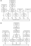
(A) Flow chart of static adhesion experiments in which HUVECs were exposed to oxLDL, histamine, and/or the supernatant of oxLDL-treated monocytes or macrophages. (B) Flow chart of static and flow adhesion experiments in which HUVECs were exposed to the supernatant of oxLDL-treated macrophages and/or the supernatant of oxLDL-treated mast cells.
Fig 2
Fig 2. Concentration of TNF-α in the supernatants of untreated monocytes (“THP-1 sup”), untreated macrophages (“Macro. Sup”), 8 μg/ml oxLDL-treated monocytes (“OxLDL-THP-1 sup”), and 8 μg/ml oxLDL-treated macrophages (“OxLDL-Macro. Sup”), according to ELISA.
Mean ± SD of three independent tests.
Fig 3
Fig 3. Number of adherent THP-1 cells on HUVEC activated by TNF-α with a concentration of 250, 500, 1000, 2000, 4000, or 10000 pg/ml.
HUVEC were exposed to TNF-α for five hours. Mean ± SD per image field (904 μm × 675 μm) of three independent tests.
Fig 4
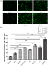
Fig 4. THP-1 cell adhesion to HUVEC exposed to oxLDL, histamine, or the macrophage supernatant.
(A) Fluorescent images of adherent THP-1 cells (green) on resting (control) or activated HUVEC. (B) Number of adherent THP-1 cells (mean ± SD, n = 4) on resting or activated HUVEC per image field (904 μm × 675 μm). The HUVEC activation groups are: 1) “Control”—RPMI-1640 medium alone; 2) “Hist”—RPMI-1640 medium with 10-6 mol/l histamine for four hours; 3) “OxLDL”- 80 μg/ml of oxLDL for 20 hours; 4) “OxLDL + Hist”- 80 μg/ml of oxLDL for 20 hours with 10-6 mol/l histamine added 4 hours before the end of the incubation time; 5) “ST“—the supernatant of 8 μg/ml oxLDL-treated THP-1 cells for five hours; 6) “ST + Hist”—ST for five hours with 10-6 mol/l histamine added four hours before the end of the incubation time; 7) “SM“—the supernatant of 8 μg/ml oxLDL-treated THP-1 macrophages for five hours; and 8) “SM + Hist”—SM for five hours with 10-6 mol/l histamine added four hours before the end of the incubation time.
Fig 5
Fig 5. Surface expression of adhesion molecules on resting HUVEC (“Control”) and on HUVEC exposed to 10-6 mol/l histamine (“Hist”), the supernatant of 8 μg/ml oxLDL-treated macrophages (“SM”), or a combination of the oxLDL-treated macrophage supernatant and histamine (“SM+Hist”).
(A) Histogram of the expression and (B) fluorescence intensity relative to the isotype control (mean ± SD, n = 3) of ICAM-1, VCAM-1 and E-selectin. (C) Percentage of HUVEC with positive expression of these adhesion molecules (mean ± SD, n = 3). Details about the HUVEC activation groups are given in Figs. 1 and 4.
Fig 6
Fig 6. Concentration of histamine in the supernatants of untreated mast cells (“Ctrl”), 8 μg/ml oxLDL-treated mast cells (“OxLDL(8 μg/ml)”), 25 μg/ml oxLDL-treated mast cells (“OxLDL(25 μg/ml)”), and mast cells exposed to the supernatant of 8 μg/ml oxLDL-treated monocytes, according to ELISA.
Mean ± SD of three independent tests.
Fig 7
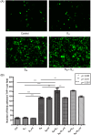
Fig 7. THP-1 cell adhesion to HUVEC exposed to the supernatants of oxLDL-treated macrophages and mast cells.
(A) Fluorescent images of adherent THP-1 cells (green) on resting (control) and activated HUVEC. (B) Number of adherent THP-1 cells (mean ± SD, n = 4) on HUVEC per image field (904 μm × 675 μm). The HUVEC activation groups are: 1) “Control”—RPMI-1640 medium alone; 2) “SL1“—the supernatant of 8 μg/ml oxLDL-treated mast cells for two hours; 3) “SL1 + H-“—SL1 combined with the antagonists of histamine receptors for two hours; 4) “SM“—the supernatant of 8 μg/ml oxLDL-treated macrophages for five hours; 5) “SM + H-“—SM combined with the antagonists of histamine receptors for five hours; 6) “SM + SL1“—SM for five hours with SL1 added two hours before the end of the incubation time; 7) “SM + SL1 + H-“—SM combined with the antagonists of histamine receptors for five hours and with SL1 added two hours before the end of the incubation time; 8) “SM + SL2“—SM for five hours with the supernatant of 25 μg/ml oxLDL-treated mast cells (SL2) added two hours before the end of the incubation time; and 9) SM + SL2 + H-—SM combined with the antagonists of histamine receptors for five hours and with SL2 added two hours before the end of the incubation time.
Fig 8
Fig 8. Number of firmly adherent THP-1 cells on resting or activated HUVEC under shear flow conditions, according to micrfluidic channel-based detachment assays.
Here, “Control”—resting endothelium, “SL1“—the supernatant of 8 μg/ml oxLDL-treated mast cells, “SM“—the supernatant of 8 μg/ml oxLDL-treated macrophages, and “SM + SL1“—the combination of the supernatants of the oxLDL-treated macrophages and mast cells. Mean μ SD of three independent tests. More details about the HUVEC activation groups are given in Figs. 1 and 7.
Fig 9
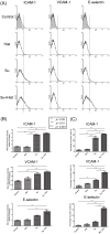
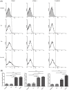
Fig 9. Surface expression of adhesion molecules on resting HUVEC (“Control”) and on HUVEC exposed to the supernatant of 8 μg/ml oxLDL-treated mast cells (“SL1”), the supernatant of 8 μg/ml oxLDL-treated macrophages (“SM”), or a combination of these supernatants (SM + SL1).
(A) Histogram of the expression of ICAM-1, VCAM-1 and E-selectin on the HUVEC surface; (B) Percentage of HUVEC with positive expression of these adhesion molecules (mean ± SD, n = 3). * p < 0.05, ** p < 0.01, *** p < 0.001. Details about the HUVEC activation groups are given in Figs. 1 and 7.

References

    1. Heart Disease & Stroke Statistics 2010 update. American Heart Association.
    1. Atherosclerosis is an inflammatory disease. Am Heart J 138: S419–420. - PubMed
    1. Moore KJ, Tabas I (2011) Macrophages in the pathogenesis of atherosclerosis. Cell 145: 341–355. 10.1016/j.cell.2011.04.005 - DOI - PMC - PubMed
    1. Badimon L, Vilahur G (2012) LDL-cholesterol versus HDL-cholesterol in the atherosclerotic plaque: inflammatory resolution versus thrombotic chaos. Ann N Y Acad Sci 1254: 18–32. 10.1111/j.1749-6632.2012.06480.x - DOI - PubMed
    1. Steinberg D (2002) Atherogenesis in perspective: hypercholesterolemia and inflammation as partners in crime. Nat Med 8: 1211–1217. - PubMed

Publication types

MeSH terms