Review
. 2015 Jul;25(7):398-407.
doi: 10.1016/j.tcb.2015.02.007. Epub 2015 Mar 24.

Mechanisms of epithelial wound detection

Affiliations
Review

Mechanisms of epithelial wound detection

Balázs Enyedi et al. Trends Cell Biol. 2015 Jul.

Abstract

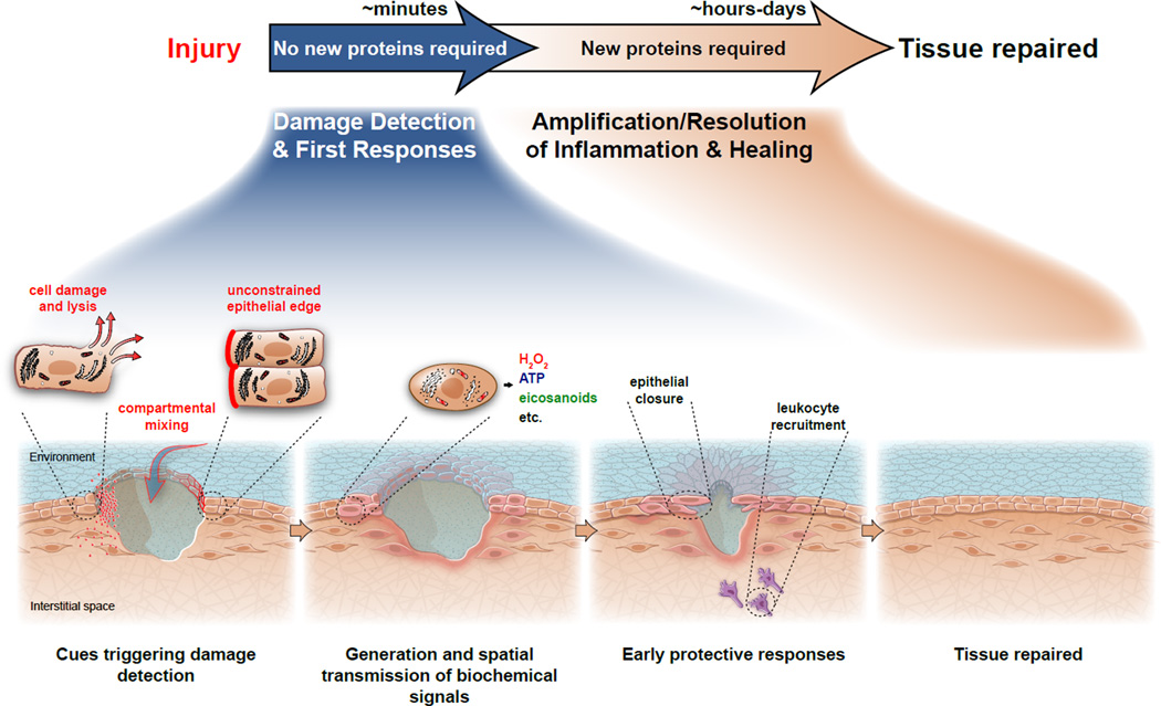
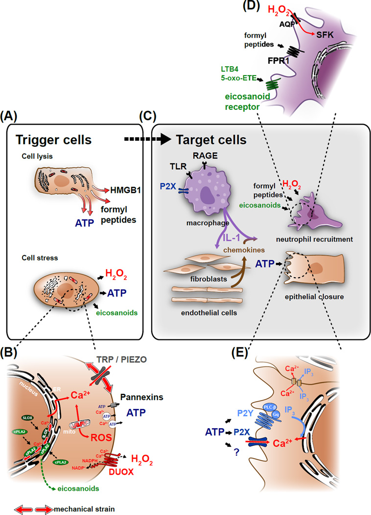
Efficient wound healing requires the coordinated responses of various cell types within an injured tissue. To react to the presence of a wound, cells have to first detect it. Judging from their initial biochemical and morphological responses, many cells including leukocytes, epithelial cells, and endothelial cells detect wounds from over hundreds of micrometers within seconds-to-minutes. Wound detection involves the conversion of an injury-induced homeostatic perturbation, such as cell lysis, an unconstrained epithelial edge, or permeability barrier breakdown, into a chemical or physical signal. The signal is spatially propagated through the tissue to synchronize protective responses of cells near the wound site and at a distance. This review summarizes the triggers and mechanisms of wound detection in animals.


Figures

Figure 1
Figure 1. Phases of wound detection and repair in the larval zebrafish
Wound detection is triggered by tissue intrinsic cues, such as factors released from lysing cells and signaling initiated in cells at unconstrained wound margins. Injury also disrupts the epithelial barrier, which leads to compartmental mixing and induces stress signaling. Minutes following injury, these signals provide spatial and temporal guidance cues for the earliest protective responses, such as epithelial closure and leukocyte recruitment. The initial ‘detection phase’ is followed by the amplification and resolution of the inflammatory response, cell proliferation, and regenerative events. These later processes are thought to be coordinated by complex transcriptional chemokine, cytokine, and growth factor cascades. Completion of tissue healing can take anywhere from hours to days.
Figure 2
Figure 2. Molecular mechanism of cell lysis- and stress-mediated damage detection
(A) Wound detection is triggered when damaged cells release cytoplasmic factors into the extracellular space. These “damage associated molecular patterns” (DAMPs) include molecules, such as HMGB1, formyl peptides or ATP. Epithelial injury also perturbs normal tissue homeostasis and results in cell stress, which induces the production of wound signals. (B) Homeostatic perturbations resulting in cell stress induce cytoplasmic Ca2+ signals by opening mechanosensitive, cation permeable channels (TRPs, Piezo) or by releasing Ca2+ from intracellular stores. Calcium signals initiate ROS production, either by activating the epithelial NADPH-oxidase DUOX (dual oxidase) through binding to the enzyme’s EF-hand domains, or by triggering mitochondrial permeability transition. Calcium signals also initiate the production and enzymatic oxidation of arachidonic acid into eicosanoids by activating cytosolic phospholipase A2 (cPLA2), and 5-lipoxygenase (5LOX). Ca2+ binding is required for these proteins to efficiently associate with nuclear membranes, giving them access to their lipid substrates. This leads to the production of proinflammatory eicosanoids, such as 5-oxo-ETE or leukotriene B4 (LTB4). Ca2+ also governs the secretion of paracrine mediators into the extracellular space (e.g., ATP) through regulating exocytosis and/or plasma membrane channel opening. (C) Most DAMPs act through transcriptional relay mechanisms in “sentinel” cells such as tissue-resident macrophages dispersed throughout the tissue. DAMPs are recognized by pattern recognition receptors (PRRs) such as toll-like receptors (TLRs), or receptors for advanced glycation end-products (RAGE). PRR and inflammasome signaling lead to processing and release of IL-1, which in turn stimulates chemokine secretion in target cells. Signals from lysed and stressed cells along with these chemokines trigger neutrophil recruitment and epithelial migration. (D) Rapid leukocyte migration to injury sites is mediated by G-protein coupled receptors, such as the N-formyl peptide receptor FPR1 and the eicosanoid receptors OXER and LTB4R. Leukocyte recruitment also depends on the H2O2-mediated oxidation of Src family kinases (SFK), such as LYN. (E) Extracellular ATP regulates wound closure by acting either as a DAMP or a stress signal through purinergic P2X channels or G-protein coupled P2Y receptors, which cause cytosolic [Ca2+]-elevation and IP3 production. Alternatively, ATP may initiate wound responses through yet unknown mechanisms.

References

    1. Szpaderska AM, et al. Differential injury responses in oral mucosal and cutaneous wounds. J. Dent. Res. 2003;82:621–626. - PubMed
    1. Seifert AW, et al. Skin regeneration in adult axolotls: a blueprint for scar-free healing in vertebrates. PLoS One. 2012;7:e32875. - PMC - PubMed
    1. Richardson R, et al. Adult zebrafish as a model system for cutaneous wound-healing research. J. Invest. Dermatol. 2013;133:1655–1665. - PMC - PubMed
    1. Gault WJ, et al. Osmotic surveillance mediates rapid wound closure through nucleotide release. J. Cell Biol. 2014;207:767–782. - PMC - PubMed
    1. Rock KL, et al. The sterile inflammatory response. Annu. Rev. Immunol. 2010;28:321–342. - PMC - PubMed

Publication types

LinkOut - more resources