Review
. 2015 Mar;39(2):105-12.
doi: 10.1053/j.semperi.2015.01.003.

Mapping the developing human brain in utero using quantitative MR imaging techniques

Affiliations
Review

Mapping the developing human brain in utero using quantitative MR imaging techniques

Colin Studholme. Semin Perinatol. 2015 Mar.

Abstract

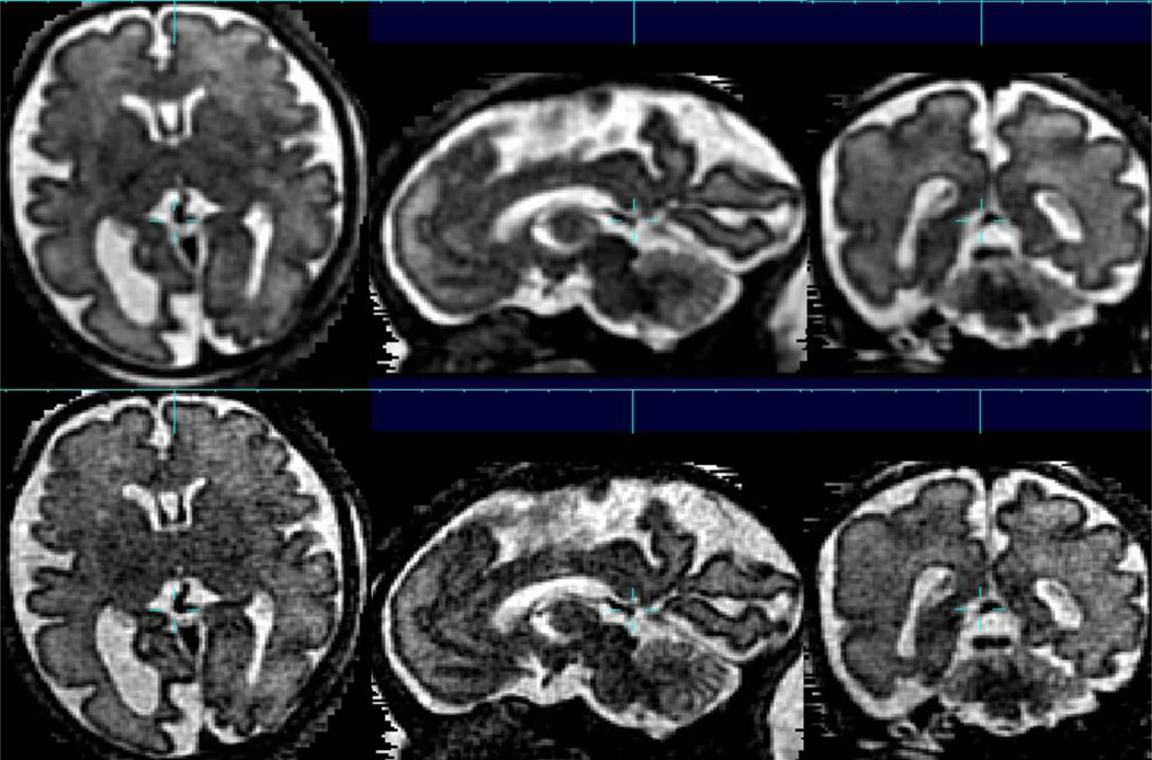
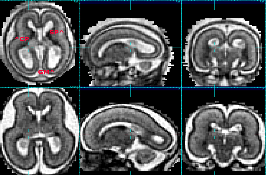
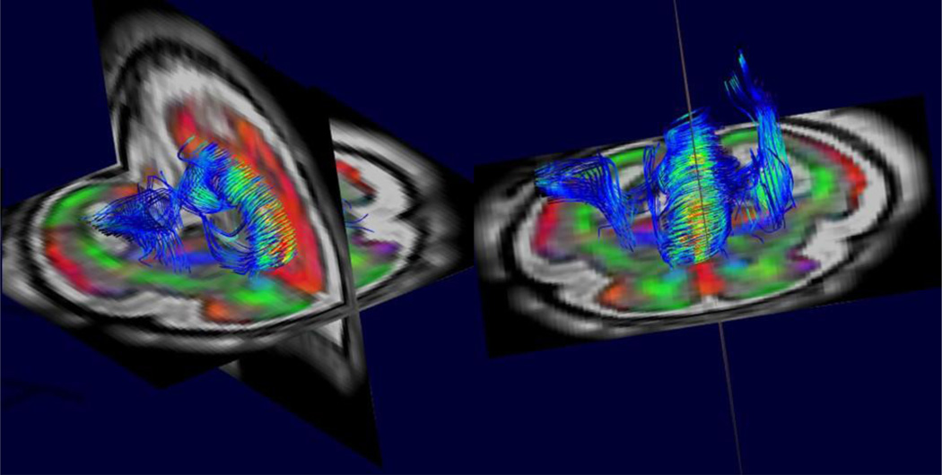
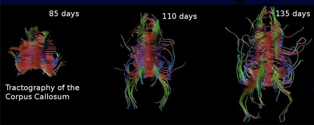
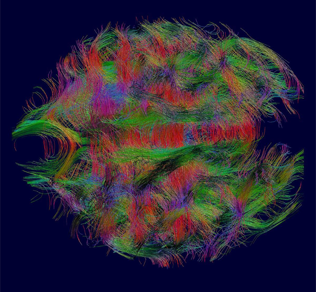
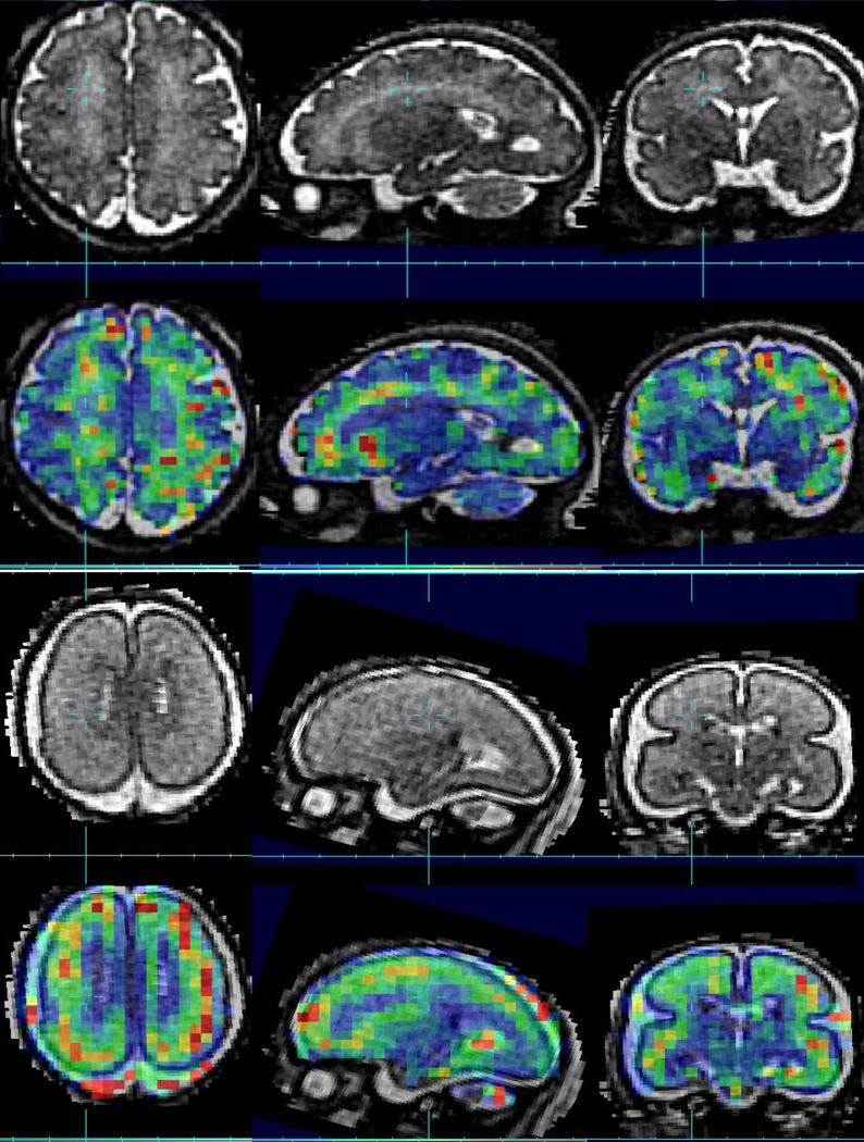
Magnetic resonance imaging of the human fetal brain has been a clinical tool for many years and provides valuable additional information to compliment more common ultrasound studies. Advances in both MRI acquisition and post processing over the last 10 years have enabled full 3D imaging and the accurate combination of data acquired in different head positions to create improved geometric integrity, tissue contrast, and resolution. This research is now motivating the development of new quantitative MRI-based techniques for clinical imaging that can more accurately characterize brain development and detect abnormalities. In this article, we will review some of the key areas that are driving changes in our understanding of fetal brain growth using quantitative measures derived from in utero MRI and the possible directions for its increased use in improving the evaluation of pregnancies and the accurate characterization of abnormal brain growth.


Figures

Figure 1
Figure 1
An example of the advantages of between slice motion correction combined with deconvolution based 3D image reconstruction (lower row) from multi-slice data, allowing a voxel size of 0.75×0.75×0.75mm to be formed from multislice, multiplane acquisitions with original slice thickness of 3.3mm. For comparison, a direct interpolation based estimated is shown in upper row. Such increased spatial resolution is particularly important in studying the cortex in later stages of gestation where the brain is becoming highly folded and complex, with many of the gyri and sulci already formed, while the brain remains relatively small in relation to its final adult size.
Figure 2
Figure 2
An example of a motion correction 3D image of conventional T2W contrast (motion corrected 1.0 × 1.0 × 1.0mm HASTE imaging) at 20GW and 23GW showing changes in contrast of developmental zones (CP=Cortical Plate, GM=Germinal Matrix, SP=Subplate) over a short time interval with sub-plate visible in the brain at 20GW loosing contrast over the next 3 or so weeks.
Figure 3
Figure 3
An example of between slice motion corrected high resolution 3D diffusion tensor imaging from combined multi-slice and multi-plane DWI. This allows tractography based analysis to quantify the development of white matter connections within the fetal brain based on the estimates of water diffusion over the brain. Note also the color coded direction map of the surrounding anatomy that highlights the highly radial structure of the cortex at this early stage of development. This contrasts with the cortical microstructure of the adult cortex which provides no directional constraints on the diffusion of water..
Figure 4
Figure 4
For basic science studies of how the human brain develops, a key advantage of MRI is the ability to study normal brain anatomy over time. Here we see the development of the corpus callosum as imaged by motion corrected DTI [47] and mapped by tractography.
Figure 5
Figure 5
Global tractography of fetal white matter can be used on motion corrected whole brain diffusion imaging studies to create a summary of interconnections between cortical and subcortical grey matter. Combined with graph analysis methods, this type of data allows us to study the development of small world connectivity [49] in the developing fetus
Figure 6
Figure 6
Example Conventional T2W contrast (1.0 × 1.0 × 1.0mm HASTE imaging) with a color overlay of quantitative T*2 imaging estimates (3.0 × 3.0 × 3.0 mm dual echo EPI) in the same fetus imaged at 20GW (lower row) and 35GW (upper row)..

References

    1. Rakic P. Mode of cell migration to the superficial layers of fetal monkey neocortex. Journal of Comparative Neurology. 1972;145(1):61–83. - PubMed
    1. Kroenke CD, Van Essen DC, Inder TE, Rees S, Bretthorst GL, Neil JJ. Microstructural changes of the baboon cerebral cortex during gestational development reflected in magnetic resonance imaging diffusion anisotropy. J Neurosci. 2007 Nov;27(46):12506–12515. Available from: http://www.hubmed.org/display.cgi?uids=18003829. - PMC - PubMed
    1. Kostović I, Judaš M, Radoš M, Hrabač P. Laminar organization of the human fetal cerebrum revealed by histochemical markers and magnetic resonance imaging. Cerebral Cortex. 2002;12(5):536–544. - PubMed
    1. Huang H, Vasung L. Gaining Insight of Fetal Brain Development with Diffusion MRI and Histology. International Journal of Developmental Neuroscience. 2013 - PMC - PubMed
    1. Huang H, Xue R, Zhang J, Ren T, Richards LJ, Yarowsky P, et al. Anatomical Characterization of Human Fetal Brain Development with Diffusion Tensor Magnetic Resonance Imaging. The Journal of Neuroscience. 2009 Apr;29(13):4263–4273. - PMC - PubMed

Publication types