

New insight into scar-related ventricular tachycardia circuits in ischemic cardiomyopathy: Fat deposition after myocardial infarction on computed tomography--A pilot study
- PMID: 25814415
- PMCID: PMC4485604
- DOI: 10.1016/j.hrthm.2015.03.041
New insight into scar-related ventricular tachycardia circuits in ischemic cardiomyopathy: Fat deposition after myocardial infarction on computed tomography--A pilot study
Abstract
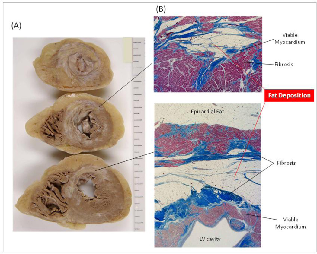
Background: Myocardial fat deposition (FAT-DEP) has been frequently observed in regions of chronic myocardial infarction in patients with ischemic cardiomyopathy. The role of FAT-DEP within scar-related ventricular tachycardia (VT) circuits has not been investigated.
Objective: This pilot study aimed to assess the impact of myocardial FAT-DEP on local electrograms and VT circuits in patients with ischemic cardiomyopathy.
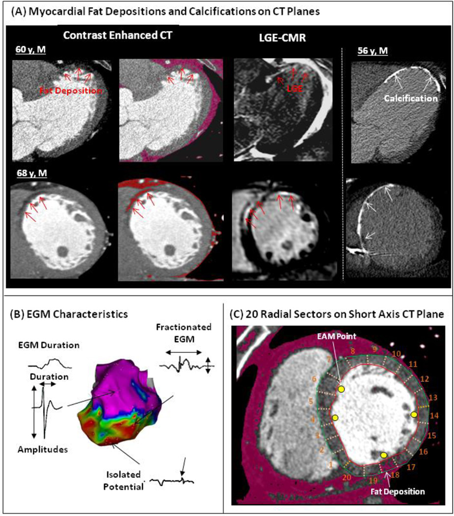
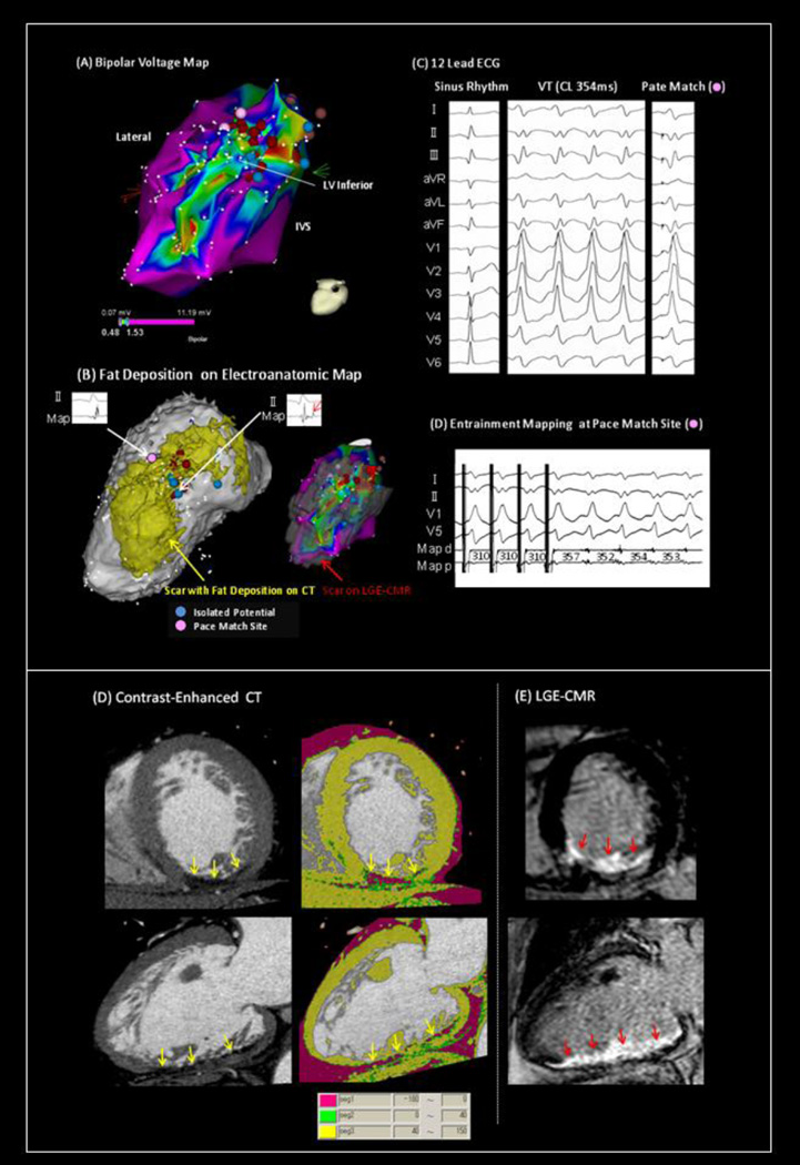
Methods: Contrast-enhanced computed tomography was performed in 22 patients with ischemic VT. Electroanatomic map points were registered to the corresponding contrast-enhanced computed tomography images. Myocardial FAT-DEP was identified and characterized using a postprocessing image overlay that highlighted areas below 0 Hounsfield units (HU). The mean attenuation of local myocardial regions corresponding to sampled electrograms was measured on short-axis images. The associations of mean attenuation with bipolar and unipolar amplitudes, left ventricular wall thickness, and VT circuit sites were investigated.
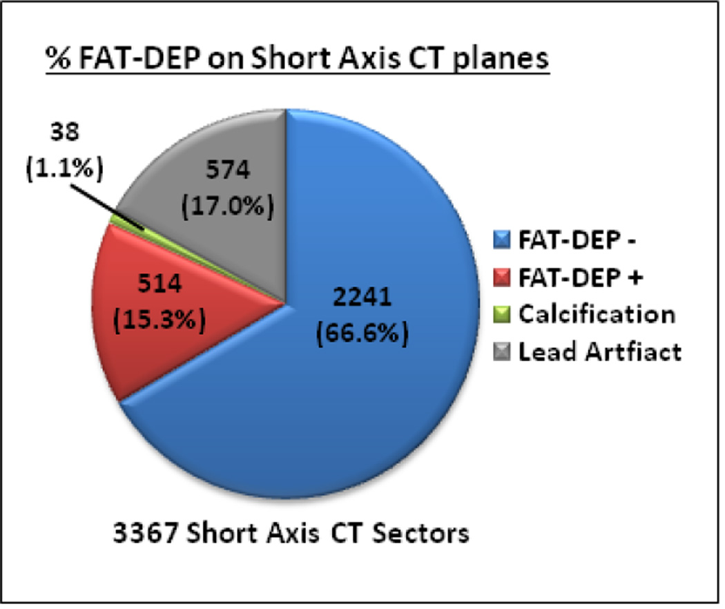
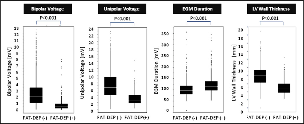
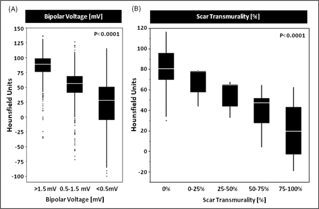
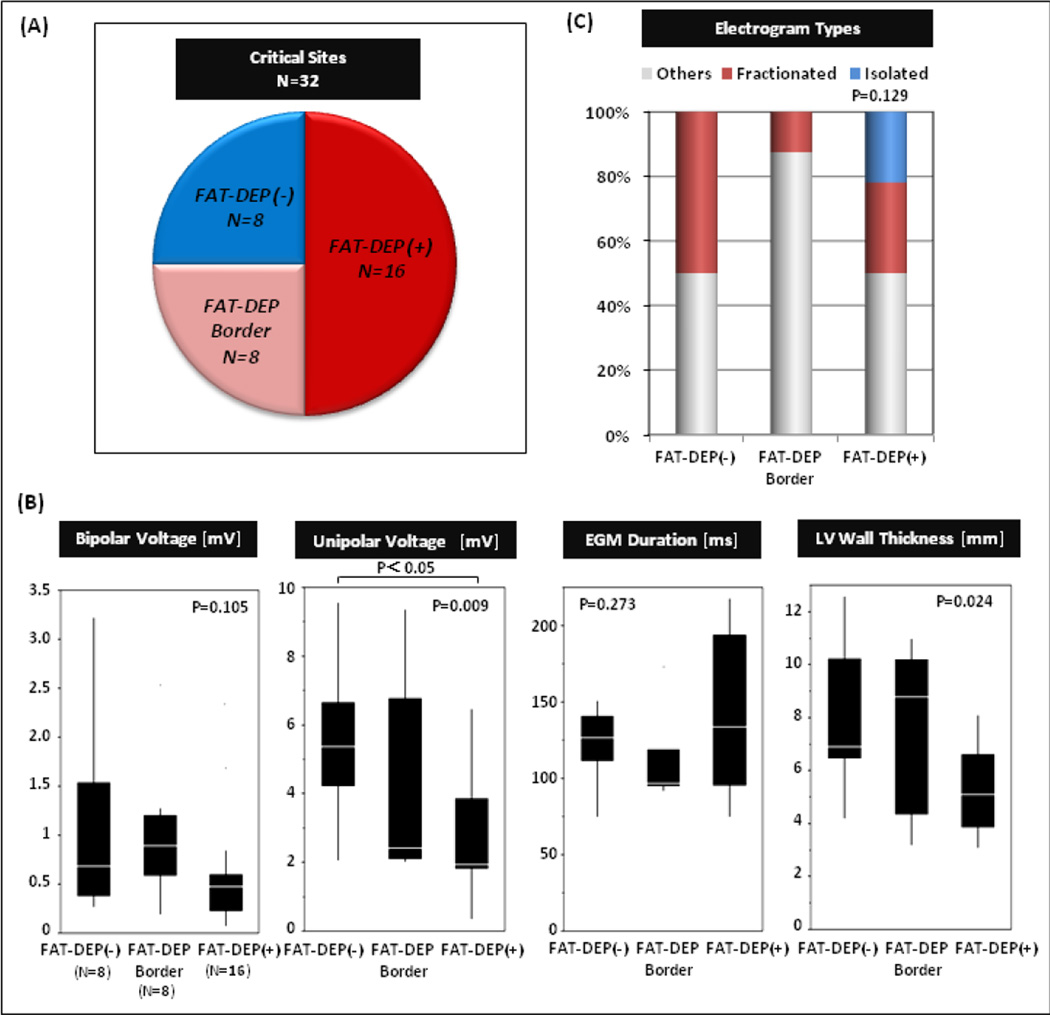
Results: Of 1801 electroanatomic map points, 519 (28.8%) were located in regions with FAT-DEP. Significant differences were observed in mean intensity (23.2 ± 35.6 HU vs 81.7 ± 21.9 HU; P < .001), bipolar (0.75 ± 0.83 mV vs 2.9 ± 2.4 mV; P < .001) and unipolar (3.1 ± 1.7 mV vs 7.4 ± 4.3 mV; P < .001) amplitudes, and left ventricular wall thickness (5.2 ± 1.7 mm vs 8.2 ± 2.5 mm; P < .001) between regions with and without FAT-DEP. Lower HU was strongly associated with lower bipolar and unipolar amplitudes (P < .0001, respectively). Importantly, FAT-DEP was associated with critical VT circuit sites with fractionated or isolated potentials.
Conclusion: FAT-DEP was associated with electrogram characteristics and VT circuit sites. Further work will be needed to determine whether FAT-DEP plays a causal role in the generation of ischemic scar-related VT circuits.
Keywords: Computed tomography; Fat; Ischemic cardiomyopathy; Magnetic resonance imaging; Ventricular tachycardia.
Copyright © 2015 Heart Rhythm Society. Published by Elsevier Inc. All rights reserved.
Conflict of interest statement
Figures
References
-
- Baroldi G, Silver MD, De Maria R, Parodi O, Pellegrini A. Lipomatous metaplasia in left ventricular scar. Can J Cardiol. 1997;13:65–71. - PubMed
-
- Su L, Siegel JE, Fishbein MC. Adipose tissue in myocardial infarction. Cardiovasc Pathol. 2004;13(2):98–102. - PubMed
-
- Winer-Muram HT, Tann M, Aisen AM, Ford L, Jennings SG, Bretz R. Computed tomography demonstration of lipomatous metaplasia of the left ventricle following myocardial infarction. J Comput Assist Tomogr. 2004;28:455–458. - PubMed
-
- Ichikawa Y, Kitagawa K, Chino S, Ishida M, Matsuoka K, Tanigawa T, Nakamura T, Hirano T, Takeda K, Sakuma H. Adipose tissue detected by multislice computed tomography in patients after myocardial infarction. JACC Cardiovasc Imaging. 2009;2:548–555. - PubMed
-
- Schmitt M, Samani N, McCann G. Images in cardiovascular medicine. Lipomatous metaplasia in Ischemic cardiomyopathy: a common but unappreciated entity. Circulation. 2007;116:e5–e6. - PubMed
Publication types
MeSH terms
Substances
Grants and funding
LinkOut - more resources
Full Text Sources
Other Literature Sources
Medical
