. 2015 Jun;8(3):516-28.
doi: 10.1161/CIRCGENETICS.114.000921. Epub 2015 Mar 26.

Nonbiased Molecular Screening Identifies Novel Molecular Regulators of Fibrogenic and Proliferative Signaling in Myxomatous Mitral Valve Disease

Affiliations

Nonbiased Molecular Screening Identifies Novel Molecular Regulators of Fibrogenic and Proliferative Signaling in Myxomatous Mitral Valve Disease

Nassir M Thalji et al. Circ Cardiovasc Genet. 2015 Jun.

Abstract

Background: Pathological processes underlying myxomatous mitral valve degeneration (MMVD) remain poorly understood. We sought to identify novel mechanisms contributing to the development of this condition.

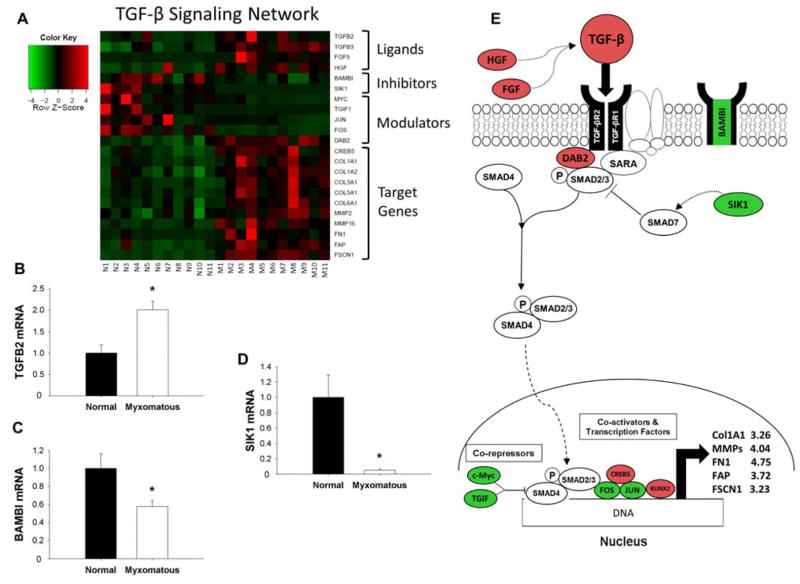
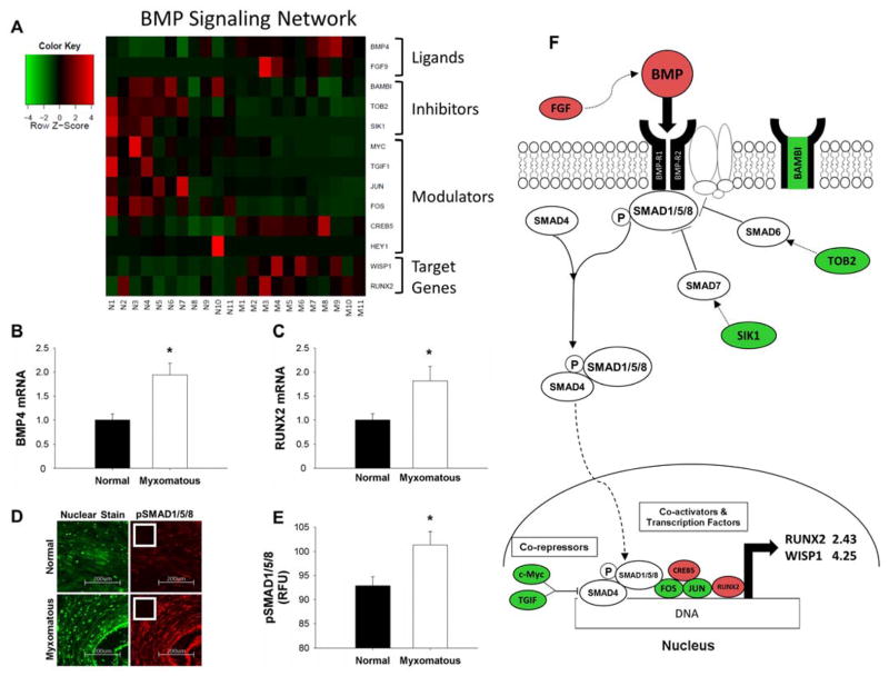
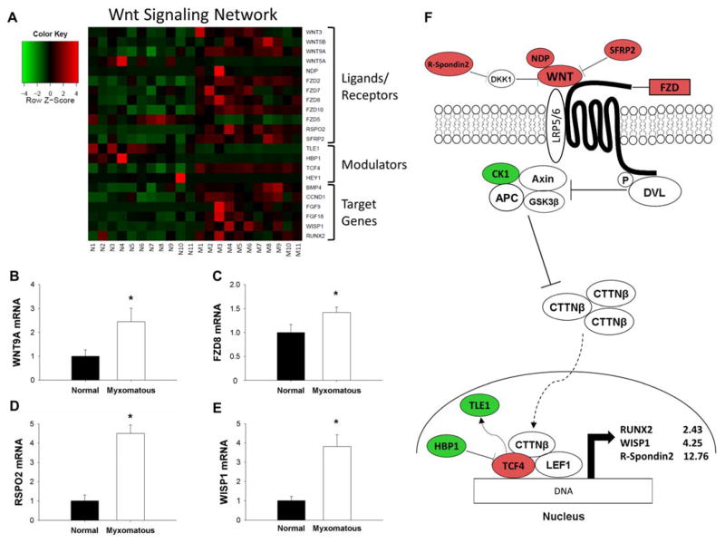
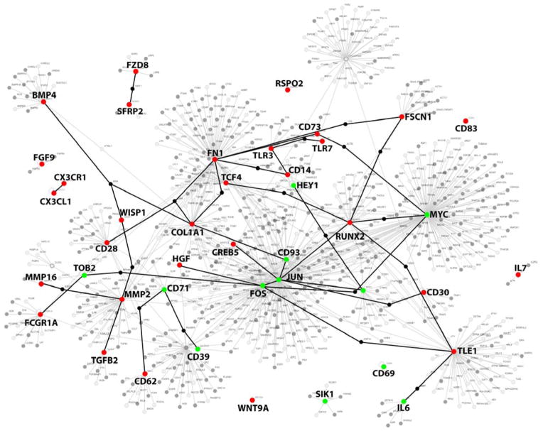
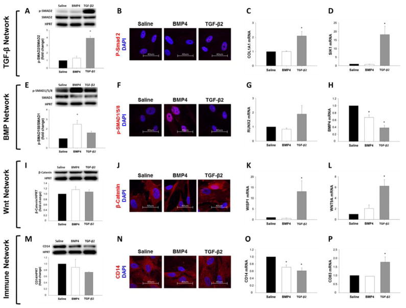
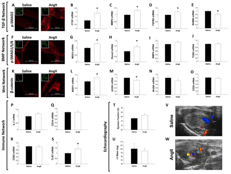
Methods and results: Microarrays were used to measure gene expression in 11 myxomatous and 11 nonmyxomatous human mitral valves. Differential gene expression (thresholds P<0.05; fold-change >1.5) and pathway activation (Ingenuity) were confirmed using quantitative reverse transcriptase polymerase chain reaction and immunohistochemistry. Contributions of bone morphogenetic protein 4 and transforming growth factor (TGF)-β2 to differential gene expression were evaluated in vitro. Contributions of angiotensin II to differential pathway activation were examined in mice in vivo. A total of 2602 genes were differentially expressed between myxomatous and nonmyxomatous valves. Canonical TGF-β signaling was increased in MMVD because of increased ligand expression and derepression of SMA mothers against decapentaplegic 2/3 signaling and was confirmed with quantitative reverse transcriptase polymerase chain reaction and immunohistochemistry. Myxomatous valves demonstrated activation of canonical bone morphogenetic protein and Wnt/β-catenin signaling and upregulation of their common target runt-related transcription factor 2. Our data set provided transcriptional and immunohistochemical evidence for activated immune cell infiltration. In vitro treatment of mitral valve interstitial cells with TGF-β2 increased β-catenin signaling at mRNA and protein levels, suggesting interactions between TGF-β2 and Wnt signaling. In vivo infusion of mice with angiotensin II recaptured several changes in signaling pathways characteristic of human MMVD.

Conclusions: These data support a new disease framework whereby activation of TGF-β2, bone morphogenetic protein 4, Wnt/β-catenin, or immune signaling plays major roles in the pathogenesis of MMVD. We propose these pathways act in a context-dependent manner to drive phenotypic changes that fundamentally differ from those observed in aortic valve disease and open novel avenues guiding future research into the pathogenesis of MMVD.

Keywords: general surgery; mitral valve; molecular biology; pathology.


Figures

Figure 1
Figure 1. Changes in Transforming Growth Factor-β (TGF-β) signaling in myxomatous mitral valve tissue
A) Heat map of differentially expressed canonical TGF-β signaling-related genes in myxomatous (columns M1–M11) versus non-myxomatous (columns N1–N11) human mitral valves. Red = increased expression, and green = reduced expression. B–D) qRT-PCR confirmation of changes in TGF-β2 ligand (B), BAMBI (C) and SIK1 (D) expression in myxomatous versus non-myxomatous mitral valve tissue (n=11 non-myxomatous valves, n=10 myxomatous valves; * = p<0.05). qRT-PCR experiments were performed on the same samples of human mitral valve tissue used for microarray analyses. E) For ease of reference, a simplified working model of alterations in TGF-β signaling in MMVD (color coding as for Panel 1A). For clarity, non-myxomatous control valves are referred to as “normal”. BAMBI = BMP and activin membrane-bound inhibitor homolog (Xenopus laevis); COL1A1 = Collagen, type I, alpha 1; COL1A2 = Collagen type I, alpha 2; COL3A1 = collagen type III, alpha 1; COL5A1 = Collagen type V, alpha 1; COL6A1 = Collagen type VI, alpha 1; CREB5 = Cycle AMP-responsive element-binding protein 5; DAB2 = Disabled homolog 2; FAP = Fibroblast activation protein; FGF9 = Fibroblast growth factor 9; FOS = FBJ murine osteosarcoma viral oncogene homolog; FN1 = Fibronectin 1; FSCN1 = Fascin 1; HGF = Hepatocyte growth factor; JUN = Jun proto-oncogene; MMP = Matrix metalloproteinase; SARA = SMAD anchor for receptor activation; SIK1 = Salt-inducible kinase 1; TGF-β2 = transforming growth factor-beta; TGF-βR = transforming growth factor-beta receptor; TGIF1 = TGF-β-induced factor homeobox 1.
Figure 2
Figure 2. Alterations in Bone Morphogenetic Protein (BMP) signaling in myxomatous mitral valves
A) Heat map of differentially expressed canonical BMP signaling-related genes in myxomatous (columns M1–M11) versus non-myxomatous (columns N1–N11) human mitral valves. Red = increased expression, green = reduced expression. qRT-PCR confirmation of changes in BMP4 ligand (B) and Runx2 (C) expression in myxomatous versus non-myxomatous mitral valves (n=11 non-myxomatous valves, n=10 myxomatous valves; * = p<0.05). qRT-PCR experiments were performed on the same samples of human mitral valve tissue used for microarray analyses. D–E) Immunohistochemical staining (D) and subsequent quantitation (E) of pSMAD1/5/8 levels in myxomatous and non-myxomatous valves (20x magnification; inset negative control; n = 11 non-myxomatous valves, n = 11 mitral valves; p < 0.05). F) For ease of reference, a simplified working model of alterations in canonical BMP signaling in MMVD (color-coding as for Panel 2A). For clarity, non-myxomatous control valves are referred to as “normal”. BAMBI = BMP and activin membrane-bound inhibitor homolog (Xenopus laevis); BMP = Bone morphogenetic protein; BMP-R = Bone morphogenetic protein receptor; CREB5 = Cycle AMP-responsive element-binding protein 5; FGF9 = Fibroblast growth factor 9; FOS = FBJ murine osteosarcoma viral oncogene homolog; HEY1 = Hairy/enhancer-of-split related with YRPW motif 1; JUN = Jun proto-oncogene; RUNX2 = Runt-related transcription factor 2; SIK1 = Salt-inducible kinase 1; SMAD = SMA mothers against decapentaplegic; TGIF1= TGF-β-induced factor homeobox1 TOB2 = Transducer of ERBB2 2; WISP1 = Wnt-inducible signaling pathway protein 1.
Figure 3
Figure 3. Changes in the Wnt/β-catenin signaling in MMVD
A) Heat map of differentially expressed canonical Wnt/β-catenin signaling-related genes in myxomatous (columns M1–M11) versus non-myxomatous (columns N1–N11) human mitral valves. Red = increased expression, green = reduced expression. B–E) qRT-PCR confirmation of differential gene expression of Wnt9a ligand (B), FZD8 receptor (C), R-spondin 2 (D) and WISP1 (E) in myxomatous versus non-myxomatous mitral valves (n = 11 non-myxomatous valves, n = 10 myxomatous valves;* = p<0.05). qRT-PCR experiments were performed on the same samples of human mitral valve tissue used for microarray analyses. F) For ease of reference, a simplified working model of canonical Wnt/β-catenin signaling in MMVD (color coding as for Panel 3A). For clarity, non-myxomatous control valves are referred to as “normal”. APC = Adenomatous polyposis coli; AXIN = Axis inhibition protein; BMP4 = bone morphogenetic protein 4; CK1 = Casein kinase 1, alpha 1; CTTNβ = Beta-catenin; DKK1 = Dickkopf 1 (Xenopus laevis); DVL = Dishevelled; FGF = fibroblast growth factor; FZD = Frizzled family receptor; HBP1 = HMG-box transcription factor 1; LEF1 = Lymphoid enhancer-binding factor 1; NDP = Norrie disease (pseudoglioma); RSPO2 = R-spondin 2; RUNX2 = Runt-related transcription factor 2; SFRP2 = Secreted frizzled-related protein 2; TCF4 = Transcription factor ; TLE1 = Transducin-like enhancer of split 1; WISP1 = Wnt-inducible signaling pathway protein 1; WNT = Wingless-type MMTV integration site family.
Figure 4
Figure 4. Changes in expression of immune cell markers and cytokines in MMVD
A) Heat map of differentially expressed genes associated with immune cell infiltration and inflammation in myxomatous (columns M1–M11) versus non-myxomatous (columns N1–N11) human mitral valves. Red = increased expression, green = reduced expression. B–D) Confirmation of changes in CD14 (B), CD83 (C), and CX3CR1 (D) gene expression in myxomatous versus non-myxomatous mitral valves (n = 11 non-myxomatous valves, n = 10 myxomatous valves; * = p<0.05). qRT-PCR experiments were performed on the same samples of human mitral valve tissue used for microarray analyses. E) Immunohistochemical staining for CD14 (20x magnification; inset negative control) in myxomatous and non-myxomatous mitral valve tissue (representative images from 11 non-myxomatous and 11 myxomatous mitral valves). F) For ease of reference, a simplified working model of potential interactions between immune cells and resident mitral valve interstitial cells in MMVD (color coding as for Panel 4A). For clarity, non-myxomatous control valves are referred to as “normal”. APC = Antigen presenting cell; CD = Cluster of differentiation; CX3CL1 = Chemokine (C-X3-C motif) ligand 1; CX3CR1 = Chemokine (C-X3-C motif) receptor 1; FCGR1A = Fc fragment of IgG high affinity Ia receptor; IL = Interleukin; TLR = Toll-like receptor.
Figure 5
Figure 5. Potential molecular cross-talk between TGF-β, BMP, Wnt/β-catenin and Immune networks in MMVD
Highlighted nodes represent genes from all heatmaps in Figures 1–4 that were differentially expressed between myxomatous and non-myxomatous valves. Red denotes increased gene expression, green denotes reduced expression. Connections between nodes (i.e. edges) represent potential first-order interactions between 2 proteins. For clarity, genes used to induce the network are highlighted in capital text, and a subset of potential interactions is highlighted using bolded edges. BMP4, Bone morphogenetic protein; CD, cluster of differentiation; CDKN1A, Cyclin-dependent kinase 1A; COL1A1, Collagen, type I, alpha I; CREB5, Cycle AMP-responsive element-binding protein 5; CX3CL1, Chemokine (C-X3-C) motif ligand 1; CX3CR1, Chemokine (C-X3-C motif) receptor 1; DAB2, Disabled homolog 2; FGF9, fibroblast growth factor 9; FN1, fibronectin 1; FOS, FBJ murine osteosarcoma viral oncogene homolog; FSCN1, Fascin 1; FZD, Frizzled family receptor; HEY1, Hairy/enhancer-of-split related with YRPW motif 1; IL6, interleukin 6; IL7, interleukin 7; JUN, Jun proto-oncogene; MMP, matrix metalloproteinase; RUNX2, Runt-related transcription factor 2; SFRP2, Secreted frizzled-related protein 2; SIK1, salt-inducible kinase 1; TCF, Transcription factor; TGFB, Transforming growth factor-β; TLE1, Transducin-like enhancer of split 1; TLR, toll-like receptor; TOB2, Transducer of ERBB2; WISP1, Wnt-inducible signaling pathway protein 1.
Figure 6
Figure 6. Effect of in vitro TGF-β and BMP4 treatment on TGF-β (A–D), BMP- (E–H), Wnt- (I–L) and immune signaling (M–P) in human mitral valve interstitial cells
pSMAD2 protein levels by western blot (A) were unchanged by BMP4 treatment but increased by TGF-β2 (24hr treatment). Immunocytochemistry (B) could not demonstrate a change in pSMAD2 with either treatment condition(100x magnification). COL1A1 (C) and SIK1 (D) gene expression levels in MVICs were not altered by BMP4 but increased by TGF-β2. pSMAD1/5/8 protein levels by western blot (E) and immunocytochemistry (100x magnification) (F) were increased by BMP4 treatment, but unaltered by TGF-β2 (24hr treatment). RUNX2 gene expression (G) by qRT-PCR was not changed by BMP4, but it tended to increase – though not significantly – following TGF-β2. BMP4 ligand levels (H) were markedly reduced by both treatments. Western blot analysis of total β-catenin protein (I) demonstrated no alteration following exogenous BMP4 or TGF-β2 (24hr treatment). Immunocytochemistry (100x magnification) (J) showed increased nuclear β-catenin with TGF-β2 but not BMP4 treatment. Gene expression of WISP1 (K) and Wnt9A (L) was unchanged by BMP4, but increased by TGF-β2. Protein levels of CD14 by western blot (M) and immunocytochemistry (100x magnification) (N) was not changed by BMP4 or TGF-β2 treatment (24hr treatment). Gene expression of CD14 by qRT-PCR (O) was reduced by both treatment conditions, while CD83 expression (P) though unaltered by BMP4 was increased by TGF-β2. BMP = Bone morphogenetic protein; CD = Cluster of differentiation; COL1A1 = collagen, type I, alpha I; Runx2 = Runt-related transcription factor 2; SIK1 = Salt-inducible kinase 1; SMAD = SMA mothers against decapentaplegic; TGF-β = Transforming growth factor-β; WISP1 = Wnt-inducible signaling pathway protein 1; Wnt9A = Wingless-type MMTV integration site family.
Figure 7
Figure 7. Molecular and phenotypic consequences of in vivo AngII infusion on murine mitral valves
A–E) Impact of AngII infusion on TGF-β signaling. Immunofluorescence of pSMAD2 (100x magnification; inset DAPI nuclear stain) (A) was enhanced in AngII treated mice. Expression of CTGF (B) and MMP2 (C), and TGF-β2 (D) was increased following AngII infusion, whereas expression of BAMBI (E) was reduced. F–J) Changes in BMP signaling following AngII treatment. Immunofluorescence of pSMAD1/5/8 (20x magnification; inset DAPI nuclear stain) (F) was unaltered by AngII infusion. mRNA levels of MSX2 (G) and Runx2 (H) were increased by AngII treatment, but expression of BMP4 (I) and TOB2 (J) was unchanged. K–O) Effects of AngII infusion on Wnt/β-catenin signaling in murine mitral valves. β-catenin immunofluorescence (20x magnification; inset DAPI nuclear stain) (K) was increased following AngII treatment. AngII increased expression of WISP1 (L) and decreased AXIN2 (M) expression. Wnt9A ligand (N) and FZD8 receptor (O) mRNA levels were unchanged by AngII infusion. P–S) Effect of AngII treatment on immune-network activation. Expression of interleukin 7 (P), CD14 (Q) and CD83 (R) was unchanged by AngII, whereas TLR7 levels (S) were increased. T–W) Echocardiographic evaluation of mitral valve and LV function in murine mitral valves. AngII infusion did not alter ejection fraction (T) or LV mass (U) in mice. V–W) Representative modified 2-chamber views of a saline-infused mouse without mitral regurgitation (V) and an AngII-infused mouse with trace mitral regurgitation (arrow) (W). (qRT-PCR: n=15 Saline vs n=13 AngII; IHC: n=8 Saline vs. n=8 AngII; * = p<0.05). AngII = Angiotensin II; AXIN2 = Axis inhibition protein 2; BAMBI = BMP and activin membrane-bound inhibitor homolog (Xenopus laevis); BMP = Bone morphogenetic protein; CD = cluster of differentiation; CTGF = Connective tissue growth factor; FZD8 = Frizzled family receptor 8; IL-7 = Interleukin 7; LV = Left ventricular; MMP2 = Matrix metalloproteinase 2; RUNX2 = Runt-related transcription factor 2; SMAD = SMA mothers against decapentaplegic; TGF-β = Transforming growth factor-β; TLR7 = Toll-like receptor 7; TOB2 = Transducer of ERBB2; WISP1; Wnt-inducible signaling pathway protein 1; Wnt9A = Wingless-type MMTV integration site family.

References

    1. Freed LA, Levy D, Levine RA, Larson MG, Evans JC, Fuller DL, et al. Prevalence and Clinical Outcome of Mitral Valve Prolapse. N Engl J Med. 1999;341:1–7. - PubMed
    1. Guthrie RB, Edwards JE. Pathology of the Myxomatous Mitral Valve: Nature, Secondary Changes and Complications. Minn Med. 1976;59:637–647. - PubMed
    1. Pomerance A. Ballooning Deformity (Mucoid Degeneration) of Atrioventricular Valves. Br Hear J. 1969;31:343–351. - PMC - PubMed
    1. Avierinos J, Gersh BJ, Melton JI, Bailey KR, Shub C, Nishimura RA, et al. Natural History of Asymptomatic Mitral Valve Prolapse in the Community. Circulation. 2002;106:1355–1361. - PubMed
    1. Suri RM, Schaff HV, Dearani Ja, Sundt TM, Daly RC, Mullany CJ, et al. Survival advantage and improved durability of mitral repair for leaflet prolapse subsets in the current era. Ann Thorac Surg. 2006;82:819–826. - PubMed

Publication types

MeSH terms