

Primary anetoderma in a young male involving palms, soles and the scalp: rarest of the rare
- PMID: 25814749
- PMCID: PMC4372953
- DOI: 10.4103/0019-5154.152605
Primary anetoderma in a young male involving palms, soles and the scalp: rarest of the rare
Abstract
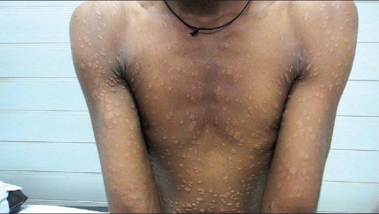
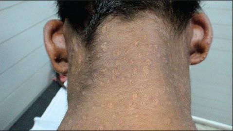
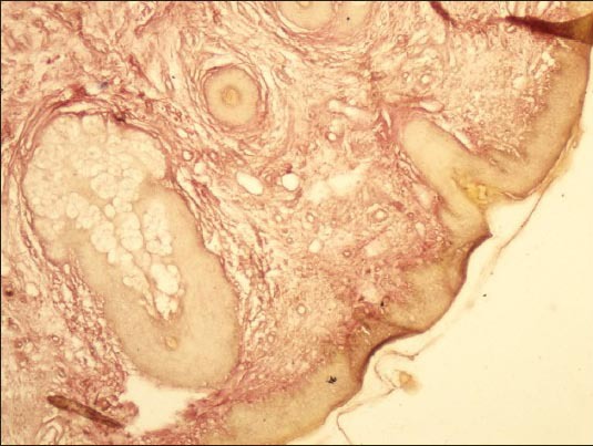
Primary anetoderma is a rare idiopathic disease of the skin, characterized by circumscribed areas of loose skin, and loss of elastic fibers upon histopathologic examination. Two forms are traditionally distinguished, primary and secondary. Primary anetoderma occurs when there is no underlying associated skin disease, whereas the latter refers to an abnormal repair mechanism of preexisting skin lesions. We are reporting a case of primary anetoderma with lesions present all over the body, including the scalp, palms and soles, the sites that are not known to be involved in this condition.
Keywords: Elastolysis and elastorrhexis; palms; primary anetoderma; soles and scalp.
Conflict of interest statement
Figures


Similar articles
-
Generalized anetoderma: An unusual manifestation of secondary syphilis treated with injection penicillin.Indian J Sex Transm Dis AIDS. 2021 Jul-Dec;42(2):166-168. doi: 10.4103/ijstd.IJSTD_81_19. Epub 2021 Jul 27. Indian J Sex Transm Dis AIDS. 2021. PMID: 34909625 Free PMC article.
-
Primary anetoderma: a cutaneous marker of antiphospholipid antibodies.Skinmed. 2011 May-Jun;9(3):168-71. Skinmed. 2011. PMID: 21675496 Review.
-
Anetoderma due to secondary syphilis: Report of two cases and discussion of the histopathological findings.Int J STD AIDS. 2017 Dec;28(14):1456-1460. doi: 10.1177/0956462417713586. Epub 2017 Jun 1. Int J STD AIDS. 2017. PMID: 28571518
-
Ultrastructural aspects of primary anetoderma.J Cutan Pathol. 2017 Sep;44(9):786-789. doi: 10.1111/cup.12978. Epub 2017 Jul 5. J Cutan Pathol. 2017. PMID: 28605051
-
Circumscribed lenticular anetoderma in an HIV-infected man with a history of syphilis and lichen planus.Dermatol Online J. 2011 Oct 15;17(10):2. Dermatol Online J. 2011. PMID: 22031628 Review.
Cited by
-
Multibacillary Leprosy Presenting as Anetoderma in a Young Teenager.Case Rep Dermatol Med. 2020 Jul 29;2020:8847310. doi: 10.1155/2020/8847310. eCollection 2020. Case Rep Dermatol Med. 2020. PMID: 32802526 Free PMC article.
References
-
- Burton JL, Lovell CR. Disorders of connective tissue. In: Champion RH, Burton JL, Burns DA, et al., editors. Textbook of dermatology. 6th ed. Vol. 3. United States: Oxford, Blackwell Science; 1998. pp. 2003–72.
-
- Venencie PY, Winkelmann RK. Histopathologic findings in anetoderma. Arch Dermatol. 1984;120:1040–4. - PubMed
-
- Venencie PY, Winkelmann RK, Moore BA. Anetoderma: Clinical findings, associations, and long term follow-up evaluations. Arch Dermatol. 1984;120:1032–9. - PubMed
-
- Staiger H, Saposnik M, Spiner RE, Schröh RG, Corbella MC, Hassan ML. Primary anetoderma and antiphospholipid antibodies. Dermatol Argent. 2008;14:372–8. - PubMed
-
- Burrows NP, Lovell CR. Disorders of Connective tissue. In: Burns T, Breathnach S, Cox N, Griffiths C, editors. Rook's Textbook of Dermatology. 8th ed. United States: Oxford, Blackwell Science; 2010. pp. 45.17–8.
LinkOut - more resources
Full Text Sources