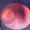
Rare Endobronchial metastasis from uterine leiomyosarcoma
- PMID: 25814801
- PMCID: PMC4372870
- DOI: 10.4103/0970-2113.152630
Rare Endobronchial metastasis from uterine leiomyosarcoma
Abstract
Uterine sarcomas are rare and represent approximately 3.2% of all invasive uterine cancers. The annual incidence rate is less than two per 100,000 women. The median age at which uterine sarcoma diagnosed is 56 years. The most common histologic pattern is leiomyosarcoma (LMS) which originates from the myometrium or myometrial vessels. Uterine LMSs are aggressive tumors with high rates of recurrence. The most common mode of spread is hematogenous, with lymphatic spread being rare. Recurrences of up to 70% are reported in stage I and II disease with the site of recurrence being distal, most commonly the lungs or the upper abdomen. But the intra bronchial spread is extremely rare. Here we are reporting a case of uterine LMS with endobronchial metastasis causing whole lung collapse.
Keywords: Endobronchial metastasis; hematogenous; leiomyosarcoma.
Conflict of interest statement
Figures


References
-
- Echt G, Jepson J, Steel J, Langholz B, Luxton G, Hernandez W, et al. Treatment of uterine sarcomas. Cancer. 1990;66:35–9. - PubMed
-
- Salazar OM, Bonfiglio TA, Patten SF, Keller BE, Feldstein M, Dunne ME, et al. Uterine sarcomas: Natural history, treatment and prognosis. Cancer. 1978;42:1152–60. - PubMed
-
- Carol L. Cancer of the corpus uteri. In: Gloeckler Ries LA, Young JL, Jr, Keel GE, Eisner MP, Lin YD, Horner MD., editors. SEER Survival Monograph: Cancer Survival Among Adults: US SEER Program, 1988-2001, Patient and Tumor Characteristics. Chapter 15. Bethesda, MD: National Cancer Institute, SEER Program, NIH; 2007. pp. 123–32.
-
- Harlow BL, Weiss NS, Lofton S. The epidemiology of sarcomas of the uterus. J Natl Cancer Inst. 1986;76:399–402. - PubMed
-
- Livi L, Paiar F, Shah N, Blake P, Villanucci A, Amunni G, et al. Uterine sarcoma: Twenty-seven years of experience. Int J Radiat Oncol Biol Phys. 2003;57:1366–73. - PubMed
LinkOut - more resources
Full Text Sources
Other Literature Sources

