Review
. 2015 Mar 12:9:80.
doi: 10.3389/fncel.2015.00080. eCollection 2015.

The choroid plexus-a multi-role player during infectious diseases of the CNS

Affiliations
Review

The choroid plexus-a multi-role player during infectious diseases of the CNS

Christian Schwerk et al. Front Cell Neurosci. .

Abstract

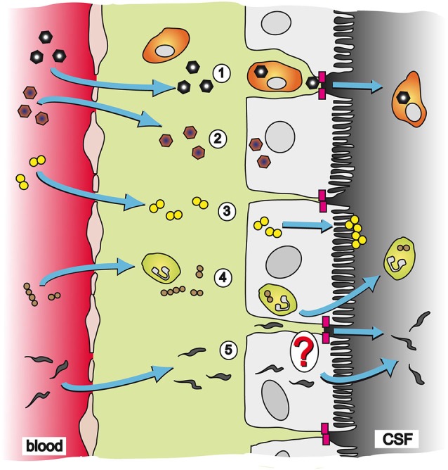
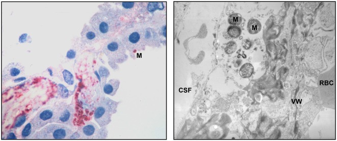
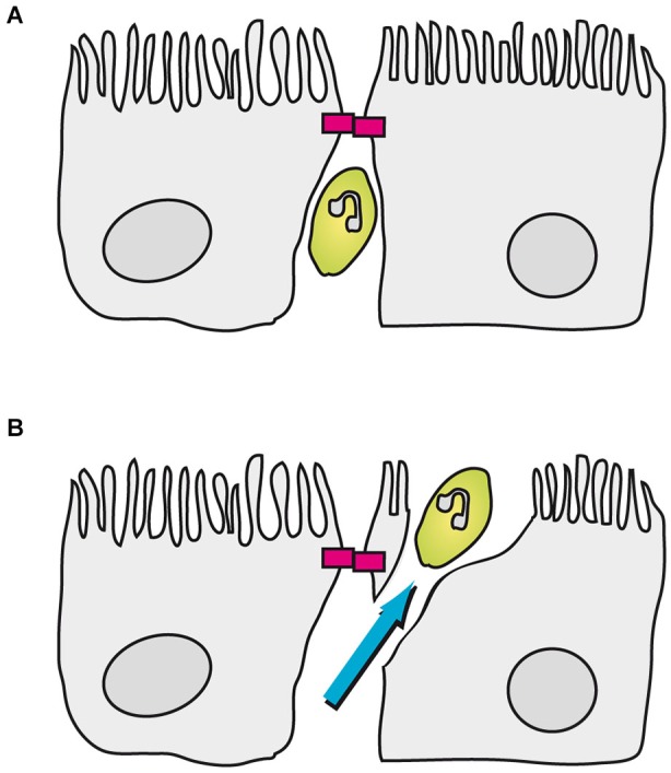
The choroid plexus (CP) is the source of cerebrospinal fluid (CSF) production and location of the blood-CSF barrier (BCSFB), which is constituted by the epithelial cells of the CP. Several infectious pathogens including viruses, bacteria, fungi and parasites cross the BCSFB to enter the central nervous system (CNS), ultimately leading to inflammatory infectious diseases like meningitis and meningoencephalitis. The CP responds to this challenge by the production of chemokines and cytokines as well as alterations of the barrier function of the BCSFB. During the course of CNS infectious disease host immune cells enter the CNS, eventually contributing to the cellular damage caused by the disease. Additional complications, which are in certain cases caused by choroid plexitis, can arise due to the response of the CP to the pathogens. In this review we will give an overview on the multiple functions of the CP during brain infections highlighting the CP as a multi-role player during infectious diseases of the CNS. In this context the importance of tools for investigation of these CP functions and a possible suitability of the CP as therapeutic target will be discussed.

Keywords: blood-brain barrier; blood-cerebrospinal fluid barrier; central nervous system infection and inflammation; choroid plexus; pathogen.


Figures

Figure 1
Figure 1
Pathways and strategies of pathogen invasion into the CNS via the CP. The CP is an endothelial-epithelial convolute responsible for production of the CSF. Whereas the blood capillaries in the CP are fenestrated, the epithelial cells of the CP are connected to each other by TJs (magenta blocks) and present the morphological correlate of the BCSFB. (1) CVB3 has been described to cross the BCSFB inside of a specific population of myeloid cells, which is highly susceptible to infection upon traversal of the CP epithelial TJs; (2) EV30 can invade into CP epithelial cells and replicate inside the host cell; (3) The bacterium N. meningitidis can directly invade into CP epithelial cells. After transmigration through the cell layer N. meningitidis forms microcolonies at the apical cell side; (4) S. suis has been shown to invade into PMNs and crosses the BCSFB in a transcellular fashion employing a “Trojan horse” strategy; and (5) The parasite T. b. brucei can transmigrate through the CP epithelium to enter the CSF. The exact mechanism of translocation by T. b. brucei is not clear yet. It is hypothesized that the parasite follows a paracellular route after opening of TJs. Alternatively, T. b. brucei could directly invade CP epithelial cells for a transcellular mechanism.
Figure 2
Figure 2
Pathologic findings in the CP of human patients with meningococcal disease. An immunohistochemical examination (left panel) shows abundant bacteria and bacterial antigens in the lumen of a thrombosed blood vessel and in the interstitial tissue. One N. meningitidis organism (M) can be seen in the surface of a CP epithelial cells. Transmission electron microscopy (right panel) demonstrates meningococci (M) in the interstitial space. The blood vessel wall (VW), a red blood cell (RBC) and the space containing cerebrospinal fluid (CSF) are pointed out. The pictures are reproduced with friendly permission from Guarner et al. (2004).
Figure 3
Figure 3
Transmigration of porcine PMNs in an in vitro model of the BCSFB based on PCPEC. In this model PMNs can be found between CP epithelial cells up to the apically located TJs (magenta blocks). (A) Once a PMN encounters a closed TJ, the neutrophil stops in front of the TJ. (B) Subsequently, the PMN finishes transmigration in a transcellular fashion, which involves the formation of a “funnel-like” structure originating from the apical membrane of the PCPEC.

References

    1. Abbott N. J. (2004). Evidence for bulk flow of brain interstitial fluid: significance for physiology and pathology. Neurochem. Int. 45, 545–552. 10.1016/j.neuint.2003.11.006 - DOI - PubMed
    1. Abbott N. J., Patabendige A. A., Dolman D. E., Yusof S. R., Begley D. J. (2010). Structure and function of the blood-brain barrier. Neurobiol. Dis. 37, 13–25. 10.1016/j.nbd.2009.07.030 - DOI - PubMed
    1. Akira S., Uematsu S., Takeuchi O. (2006). Pathogen recognition and innate immunity. Cell 124, 783–801. 10.1016/j.cell.2006.02.015 - DOI - PubMed
    1. Amasheh S., Meiri N., Gitter A. H., Schöneberg T., Mankertz J., Schulzke J. D., et al. . (2002). Claudin-2 expression induces cation-selective channels in tight junctions of epithelial cells. J. Cell Sci. 115, 4969–4976. 10.1242/jcs.00165 - DOI - PubMed
    1. Arends J. P., Zanen H. C. (1988). Meningitis caused by Streptococcus suis in humans. Rev. Infect. Dis. 10, 131–137. 10.1093/clinids/10.1.131 - DOI - PubMed

LinkOut - more resources