. 2015 Mar 12:9:84.
doi: 10.3389/fncel.2015.00084. eCollection 2015.

Brain region-specific gene expression profiles in freshly isolated rat microglia

Affiliations

Brain region-specific gene expression profiles in freshly isolated rat microglia

Karlijn J Doorn et al. Front Cell Neurosci. .

Abstract

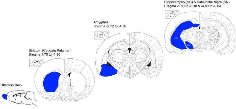
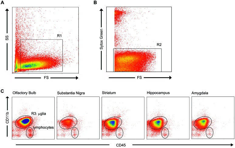
Microglia are important cells in the brain that can acquire different morphological and functional phenotypes dependent on the local situation they encounter. Knowledge on the region-specific gene signature of microglia may hold valuable clues for microglial functioning in health and disease, e.g., Parkinson's disease (PD) in which microglial phenotypes differ between affected brain regions. Therefore, we here investigated whether regional differences exist in gene expression profiles of microglia that are isolated from healthy rat brain regions relevant for PD. We used an optimized isolation protocol based on a rapid isolation of microglia from discrete rat gray matter regions using density gradients and fluorescent-activated cell sorting. Application of the present protocol followed by gene expression analysis enabled us to identify subtle differences in region-specific microglial expression profiles and show that the genetic profile of microglia already differs between different brain regions when studied under control conditions. As such, these novel findings imply that brain region-specific microglial gene expression profiles exist that may contribute to the region-specific differences in microglia responsivity during disease conditions, such as seen in, e.g., PD.

Keywords: FACS; Parkinson’s disease; hippocampus; microglia; olfactory bulb; substantia nigra.


Figures

FIGURE 1
FIGURE 1
Anatomical representation of dissected brain regions (adapted from the Rat Brain Atlas; https://gaidi.ca/rat-brain-atlas/). Brain regions that were dissected for microglia isolation are indicated in blue; OB, STR, AM, HC and SN. Bregma coordinates indicate the anterior-posterior range of each region that was dissected.
FIGURE 2
FIGURE 2
Flow cytometric analysis based on cell size, granularity and viability identified microglia with CD11bhigh/CD45pos expression. (A) Microglia appeared homogeneous in cell size and granularity (R1). (B) Sytox green staining, a DNA-binding dye that is actively taken up by dying cells, distinguished the living cells (R2) from dead cells and debris. (C) Combined selection based on cell size, granularity, and viability (R1 and R2) resulted in a microglial purity of 92.4 ± 0.87% (mean ± SD), as identified by their characteristic CD11bhigh/CD45pos expression. A small fraction of lymphocytes was present to within the viable cells. To obtain a 100% pure microglial population, microglia (R3) were sorted.
FIGURE 3
FIGURE 3
Region-specific expression levels of established microglial markers. No significant differences were found in AIF1 (A), CD11b (B) and TLR2 (C) mRNA levels between the different brain regions. For CD68 (D) and IL-1β (E), a significantly higher expression was present in the OB compared to other brain regions (CD68: OB vs. Str, HC, AM p < 0.05; IL-1β: OB vs. Str p < 0.05), and a significantly higher expression of CD68 (d) in Str compared to AM was found (#p < 0.02). Significantly higher TNF mRNA level (F) was measured in the SN relative to the HC (p < 0.05), a significantly higher CCR2 mRNA level (G) in SN compared to OB (p < 0.05), significantly lower P2X7R mRNA level (H) in SN compared to Str, HC, and AM (p < 0.05), and significantly more P2X7R expression (H) in the HC compared to OB (#p < 0.05). (I) No GFAP expression was observed, indicating that the sorted microglia were not contaminated by astrocytes.

References

    1. Aloisi F. (2001). Immune function of microglia. Glia 36 165–179 10.1002/glia.1106 - DOI - PubMed
    1. Amino T., Orimo S., Itoh Y., Takahashi A., Uchihara T., Mizusawa H. (2005). Profound cardiac sympathetic denervation occurs in Parkinson disease. Brain Pathol. 15 29–34 10.1111/j.1750-3639.2005.tb00097.x - DOI - PMC - PubMed
    1. Beraud D., Twomey M., Bloom B., Mittereder A., Ton V., Neitzke K., et al. (2011). α-Synuclein alters toll-like receptor expression. Front. Neurosci. 5:80 10.3389/fnins.2011.00080 - DOI - PMC - PubMed
    1. Borrajo A., Rodriguez-Perez A. I., Diaz-Ruiz C., Guerra M. J., Labandeira-Garcia J. L. (2014). Microglial TNFα mediates enhancement of dopaminergic degeneration by brain angiotensin. Glia 62 145–157 10.1002/glia.22595 - DOI - PubMed
    1. Braak H., Braak E. (2000). Pathoanatomy of Parkinson’s disease. J. Neurol. 247 3–10 10.1007/PL00007758 - DOI - PubMed