Case Reports
. 2015 Mar 18;4(3):2047981614560938.
doi: 10.1177/2047981614560938. eCollection 2015 Mar.

SWI enhances vein detection using gadolinium in multiple sclerosis

Affiliations
Case Reports

SWI enhances vein detection using gadolinium in multiple sclerosis

Pietro Maggi et al. Acta Radiol Open. .

Abstract

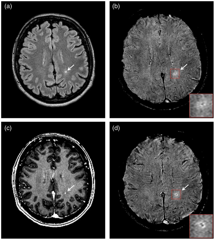
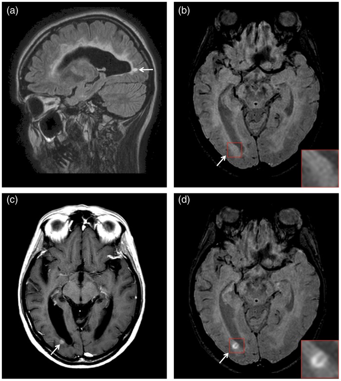
Susceptibility weighted imaging (SWI) combined with the FLAIR sequence provides the ability to depict in vivo the perivenous location of inflammatory demyelinating lesions - one of the most specific pathologic features of multiple sclerosis (MS). In addition, in MS white matter (WM) lesions, gadolinium-based contrast media (CM) can increase vein signal loss on SWI. This report focuses on two cases of WM inflammatory lesions enhancing on SWI images after CM injection. In these lesions in fact the CM increased the contrast between the parenchyma and the central vein allowing as well, in one of the two cases, the detection of a vein not visible on the same SWI sequence acquired before CM injection.

Keywords: Central nervous system (CNS); brain/brain stem; inflammation; magnetic resonance imaging (MRI).


Figures

Fig. 1.
Fig. 1.
Left peritrigonal focal WM lesion (arrows) appearing hyperintense on (a) the axial pre-CM FLAIR and (b) the axial pre-CM SWI. In addition SWI shows a small faint hypointense area in the lesion center (magnified view) that can be ascribed to the presence of a central vein. (c) The lesion appears visibly contrast-enhancing on the axial post-CM MPRAGE and (d) the axial post-CM SWI. The hypointense vein (already visible on the precontrast SWI) here appears enlarged and the contrast between the small hyperintense contrast-enhancing lesion and its hypointense central vein, appears increased (magnified view).
Fig. 2.
Fig. 2.
Occipital periventricular WM lesion (arrows) appearing hyperintense on (a) the sagittal pre-CM FLAIR and (b) the pre-CM axial SWI (magnified view). (c) The lesion appears visibly contrast-enhancing on the post-CM axial T1W SE and (d) the post-CM axial SWI. The post-CM SWI shows a linear shaped area of signal hypointensity in the lesion center, a parenchymal vein, which is not visible on the pre-CM SWI (magnified view).

References

    1. Mittal S, Wu Z, Neelavalli J, et al. Susceptibility-weighted imaging: technical aspects and clinical applications, part 2. Am J Neuroradiol 2009; 30: 232–252. - PMC - PubMed
    1. Sati P, Thomasson D, Li N, et al. Rapid, high-resolution, whole-brain, susceptibility-based MRI of multiple sclerosis. Mult Scler 2014; 20: 1464–1470. - PMC - PubMed
    1. Gaitan MI, Maggi P, Wohler J, et al. Perivenular brain lesions in a primate multiple sclerosis model at 7-tesla magnetic resonance imaging. Mult Scler 2013; 20: 64–71. - PMC - PubMed
    1. Haacke EM, Mittal S, Wu Z, et al. Susceptibility-weighted imaging: technical aspects and clinical applications, part 1. Am J Neuroradiol 2009; 30: 19–30. - PMC - PubMed
    1. Absinta M, Sati P, Gaitan MI, et al. Seven-tesla phase imaging of acute multiple sclerosis lesions: A new window into the inflammatory process. Ann Neurol 2013; 74: 669–678. - PMC - PubMed

Publication types

LinkOut - more resources