

Hematopoietic cancer cell lines can support replication of Sabin poliovirus type 1
- PMID: 25815312
- PMCID: PMC4359862
- DOI: 10.1155/2015/358462
Hematopoietic cancer cell lines can support replication of Sabin poliovirus type 1
Abstract
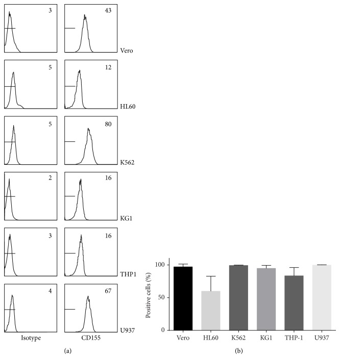
Viral vaccines can be produced in adherent or in suspension cells. The objective of this work was to screen human suspension cell lines for the capacity to support viral replication. As the first step, it was investigated whether poliovirus can replicate in such cell lines. Sabin poliovirus type 1 was serially passaged on five human cell lines, HL60, K562, KG1, THP-1, and U937. Sabin type 1 was capable of efficiently replicating in three cell lines (K562, KG1, and U937), yielding high viral titers after replication. Expression of CD155, the poliovirus receptor, did not explain susceptibility to replication, since all cell lines expressed CD155. Furthermore, we showed that passaged virus replicated more efficiently than parental virus in KG1 cells, yielding higher virus titers in the supernatant early after infection. Infection of cell lines at an MOI of 0.01 resulted in high viral titers in the supernatant at day 4. Infection of K562 with passaged Sabin type 1 in a bioreactor system yielded high viral titers in the supernatant. Altogether, these data suggest that K562, KG1, and U937 cell lines are useful for propagation of poliovirus.
Figures


Similar articles
-
Mathematical model of adherent Vero cell growth and poliovirus production in animal component free medium.Bioprocess Biosyst Eng. 2015 Mar;38(3):543-55. doi: 10.1007/s00449-014-1294-2. Epub 2014 Oct 8. Bioprocess Biosyst Eng. 2015. PMID: 25294335
-
Hematopoietic cells from CD155-transgenic mice express CD155 and support poliovirus replication ex vivo.Microb Pathog. 2000 Oct;29(4):203-12. doi: 10.1006/mpat.2000.0386. Microb Pathog. 2000. PMID: 10993739
-
K562 cell strains differ in their response to poliovirus infection.Virology. 1995 Oct 20;213(1):7-18. doi: 10.1006/viro.1995.1541. Virology. 1995. PMID: 7483280
-
One hundred years of poliovirus pathogenesis.Virology. 2006 Jan 5;344(1):9-16. doi: 10.1016/j.virol.2005.09.015. Virology. 2006. PMID: 16364730 Review.
-
[Poliovirus susceptibility in cultured cells--an answer to Enders].Uirusu. 2006 Jun;56(1):59-66. doi: 10.2222/jsv.56.59. Uirusu. 2006. PMID: 17038813 Review. Japanese.
Cited by
-
Interaction between nectin-1 and the human natural killer cell receptor CD96.PLoS One. 2019 Feb 13;14(2):e0212443. doi: 10.1371/journal.pone.0212443. eCollection 2019. PLoS One. 2019. PMID: 30759143 Free PMC article.
-
Dasatinib-induced anti-leukemia cellular immunity through a novel subset of CD57 positive helper/cytotoxic CD4 T cells in chronic myelogenous leukemia patients.Int J Hematol. 2018 Dec;108(6):588-597. doi: 10.1007/s12185-018-2517-0. Epub 2018 Aug 27. Int J Hematol. 2018. PMID: 30151740
References
-
- Hayflick L., Plotkin S. A., Norton T. W., Koprowski H. Preparation of poliovirus vaccines in a human fetal diploid cell strain. The American Journal of Epidemiology. 1962;75(2):240–258. - PubMed
-
- Zealley H., Edmond E., Inglis J. M., Shepherd W. M., Langford D. T. A comparison of the reactivity and immunogenicity of RA 27/3 strain rubella vaccine prepared in WI-38 or MRC5 human diploid cells. Journal of Biological Standardization. 1986;14(3):213–216. doi: 10.1016/0092-1157(86)90005-3. - DOI - PubMed
MeSH terms
Substances
LinkOut - more resources
Full Text Sources
Other Literature Sources
Research Materials
