. 2014 Nov 26;2(1):31-43.
doi: 10.18632/oncoscience.100. eCollection 2015.

Human RNASET2 derivatives as potential anti-angiogenic agents: actin binding sequence identification and characterization

Affiliations

Human RNASET2 derivatives as potential anti-angiogenic agents: actin binding sequence identification and characterization

Liron Nesiel-Nuttman et al. Oncoscience. .

Abstract

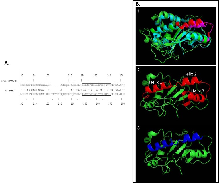
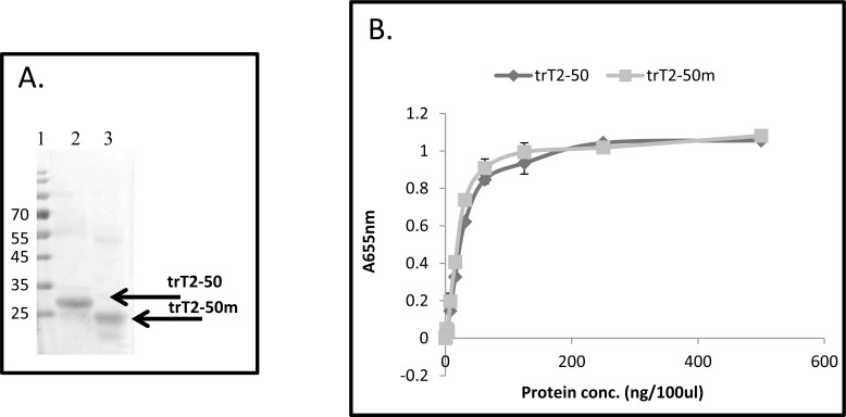
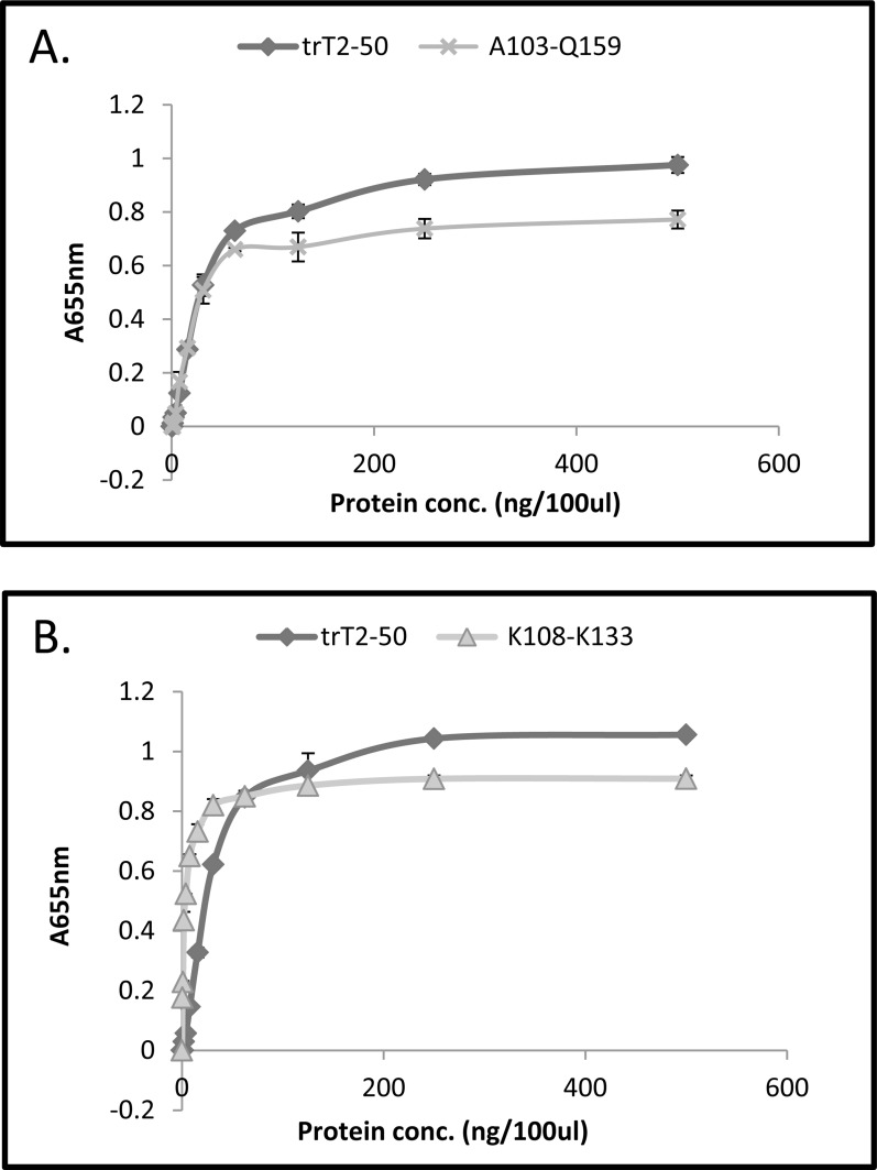
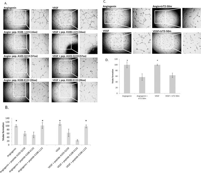
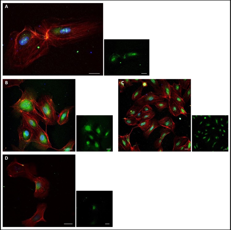
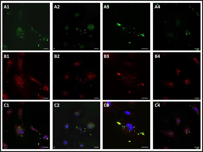
Human RNASET2 (hRNASET2) has been demonstrated to exert antiangiogenic and antitumorigenic effects independent of its ribonuclease capacity. We suggested that RNASET2 exerts its antiangiogenic and antitumorigenic activities via binding to actin and consequently inhibits cell motility. We focused herein on the identification of the actin binding site of hRNASET2 using defined sequences encountered within the whole hRNASET2 protein. For that purpose we designed 29 different hRNASET2-derived peptides. The 29 peptides were examined for their ability to bind immobilized actin. Two selected peptides-A103-Q159 consisting of 57 amino acids and peptide K108-K133 consisting of 26 amino acids were demonstrated to have the highest actin binding ability and concomitantly the most potent anti-angiogenic activity. Further analyses on the putative mechanisms associated with angiogenesis inhibition exerted by peptide K108-K133 involved its location during treatment within the HUVE cells. Peptide K108-K133 readily penetrates the cell membrane within 10 min of incubation. In addition, supplementation with angiogenin delays the entrance of peptide K108-K133 to the cell suggesting competition on the same cell internalization route. The peptide was demonstrated to co-localize with angiogenin, suggesting that both molecules bind analogous cellular epitopes, similar to our previously reported data for ACTIBIND and trT2-50.

Keywords: RNASET2; actin-binding; antiangiogenesis; peptides; ribonuclease.


Figures

Figure 1
Figure 1
(A): Sequence alignment of ACTIBIND and human RNASET2. The box represents the sequence identified by Gundampati et al. 2012 to bind actin and the homologous sequence on the human protein sequence [26]. According to this comparison, residues 120-141, and specifically residues 135-141, were identified to exclusively contribute to the binding of hRNASET2 to actin. (B): Structural analysis using PyMole (an open-source and widely used biomolecular visualization program) and based on Thorn et al. 2012 [21]. 1: ACTIBIND and the hRNASET2 structures. In green-ACTIBIND. In light blue-hRNASET2. In pink-ACTIBIND sequence identified by Gundampati et al. 2012 to bind actin (T149-L171) [26]. In red-hRNASET2 homologous sequence (E120-A141). 2: hRNASET2 structure. In red- the longest peptide consists of 57amino acids (A103-Q157) that was identified in this work to bind actin and to inhibit angiogenesis. 3: hRNASET2 structure. In blue- the shortest peptide consisting of 26 amino acids (K108-K133) that was identified in this work to bind actin and to inhibit angiogenesis.
Figure 2
Figure 2
(A): Purified trT2-50 and trT2-50m on a 12.5% SDS-PAGE. Lane 1: molecular markers. Lanes 2: purified trT2-50. Lane 3: purified trT2-50m. (B): trT2-50m binds actin on solid phase. trT2-50 or trT2-50m were added to immobilized actin at increasing concentrations from 0 to 500 ng/100μl/well, and then reacted with rabbit anti-trT2-50 and goat anti rabbit IgG-HRP. The amount of bound protein was quantified by measuring the optical density at 655nm. Optical density was elevated in correlation to trT2-50 and trT2-50m increasing concentrations.
Figure 3
Figure 3. Peptide A103-Q159 (57aa) (A) and Peptide K108-K133 (26aa) (B) binds actin on solid phase
trT2-50 or the peptides were added to immobilized actin at increasing concentrations from 0 to 500 ng/100μl/well, and then reacted with rabbit anti-trT2-50 and goat anti rabbit IgG-HRP. The amount of bound protein and peptides was quantified by measuring the optical density at 655nm. Optical density was elevated in correlation to trT2-50 and peptides increasing concentrations.
Figure 4
Figure 4. trT2-50m and peptides A103-Q159 (57aa) and K108-K133 (26aa) inhibit angiogenin- and VEGF-induced HUVEC tube formation on Matrigel
Freshly isolated HUVECs were plated in a 96-well plate previously coated with Matrigel. (A): Cells were treated with either peptide A103-Q159, K108-K133 or K108-L123 (2μM) or PBS (control), in addition to angiogenin or VEGF (1 μg/ml). (B): tube formation was assessed using Image J, the results are represented as percent of control. Peptides A103-Q159 and K108-K133 had a significant inhibitory effect on tube formation in the presence of angiogenin and VEGF. (C): Cells were treated with trT2-50m (2μM) or PBS (control), in addition to angiogenin or VEGF (1 μg/ml). (D): tube formation was assessed using Image J, the results are represented as percent of control. trT2-50m had a significant inhibitory effect on tube formation in the presence of angiogenin and VEGF. *Represents a statistically significant difference between − and +, as analyzed using one way ANOVA for multiple comparison and 2-sample t-test, P < 0.05.
Figure 5
Figure 5. Peptide K108-K133 (26aa) is localized to the cell
(A): 10 min of incubation with peptide K108-K133-peptide was located to the the cytoplasm surrounding the nucleus (green). (B): 2 h of incubation with peptide K108-K133-the peptide was accumulated in the cytoplasm surrounding the nucleus (green). (C): 8 h of incubation with peptide K108-K133-the peptide was accumulated in the cytoplasm surrounding the nucleus (green). (D): 24 h of incubation with peptide K108-K133-the peptide had minor presence inside the cell (green). Scale bar: 20μm
Figure 6
Figure 6. Peptide K108-K133 co-localize with angiogenin in HUVECs
(A): peptide K108-K133 (green). (B): anti-angiogenin (red). (C): The integration of A and B (nucleus in blue, peptide in green and angiogenin in red). (A1): 10 min of incubation with the peptide and angiogenin– most of the peptide was localized outside the cell (green). (A2-A3): 2 h of incubation with the peptide and angiogenin– some of the peptide was observed in the cytoplasm surrounding the nucleus and some was located outside the cell (green). (A4): 8 h of incubation with the peptide and angiogenin– some of the peptide was observed in the cytoplasm surrounding the nucleus and some was located outside the cell (green). (B1): anti-angiogenin, 10 min of incubation with the peptide and angiogenin-angiogenin was located in the nucleus, the cytoplasm surrounding the nucleus, on the actin fibers and outside the cell (red). Same angiogenin localization was observed after 2 and 8 h of incubation with the peptide and angiogenin (B2, B3: 2 h. B4: 8 h) (red). (C1-C4): angiogenin and peptide K108-K133 generally co-localized mainly outside the cell after 10 min (C1), 2 h (C2, C3) and 8 h (C4).

Similar articles

Cited by

References

    1. Folkman J. Biology of Endothelial Cells. Vol. 27. Boston, MA: Springer US; 1984. pp. 412–428.
    1. Sheetz MP, Felsenfeld D, Galbraith CG, Choquet D. Cell migration as a five-step cycle. Biochem Soc Symp. 1999;65:233–43. - PubMed
    1. Roiz L, Smirnoff P, Bar-Eli M, Schwartz B, Shoseyov O. ACTIBIND, an actin-binding fungal T2-RNase with antiangiogenic and anticarcinogenic characteristics. Cancer. 2006;106:2295–308. - PubMed
    1. Morales-Ruiz M, Fulton D, Sowa G, Languino LR, Fujio Y, Walsh K, Sessa WC. Vascular endothelial growth factor-stimulated actin reorganization and migration of endothelial cells is regulated via the serine/threonine kinase Akt. Circ Res. 2000;86:892–6. - PubMed
    1. Folkman J. Angiogenesis: an organizing principle for drug discovery? Nat Rev Drug Discov. 2007;6:273–86. - PubMed

LinkOut - more resources