. 2015;46(6):2317-26.
doi: 10.3892/ijo.2015.2939. Epub 2015 Mar 27.

Taurolithocholic acid promotes intrahepatic cholangiocarcinoma cell growth via muscarinic acetylcholine receptor and EGFR/ERK1/2 signaling pathway

Affiliations

Taurolithocholic acid promotes intrahepatic cholangiocarcinoma cell growth via muscarinic acetylcholine receptor and EGFR/ERK1/2 signaling pathway

Sumet Amonyingcharoen et al. Int J Oncol. 2015.

Abstract

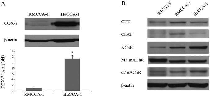
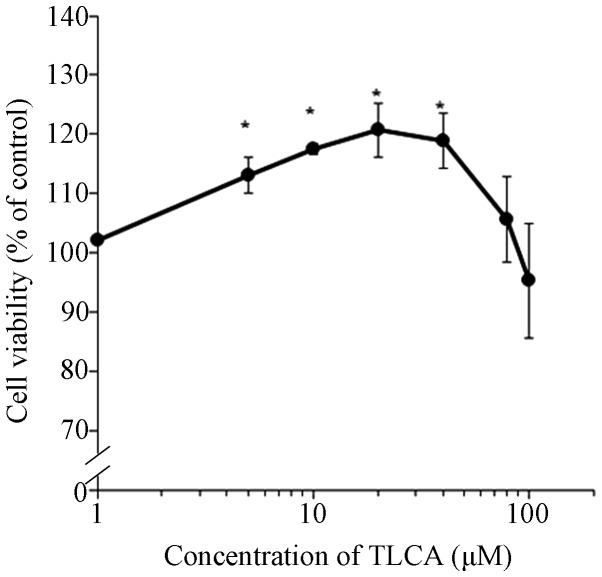
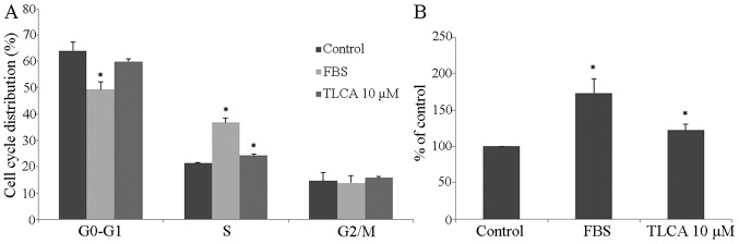
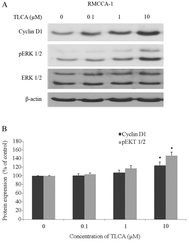
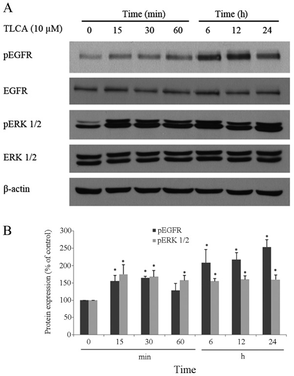
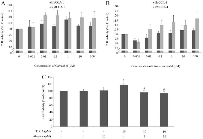
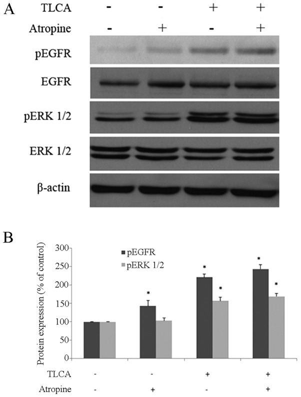
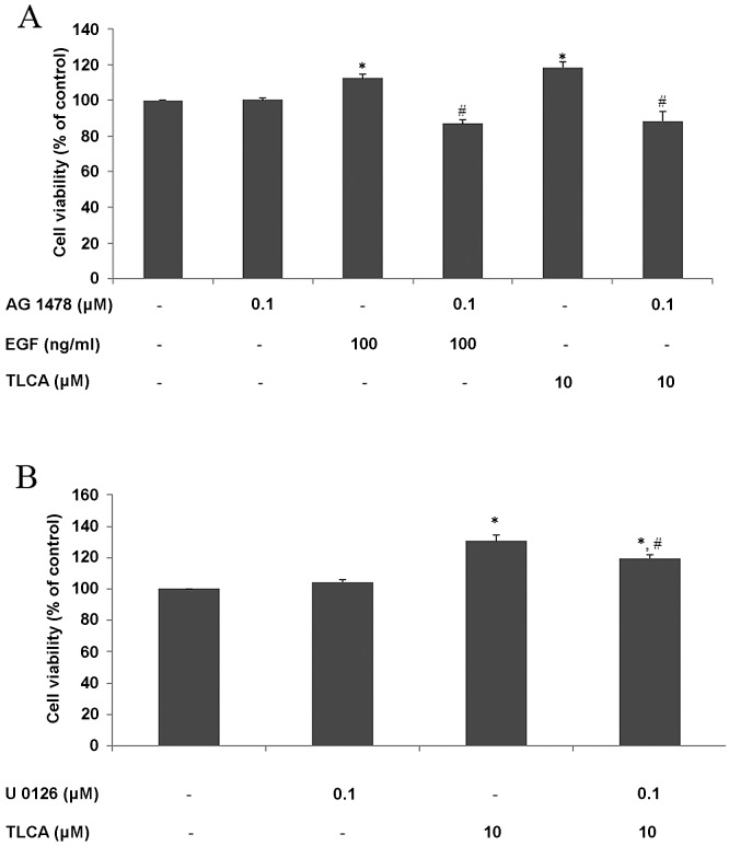
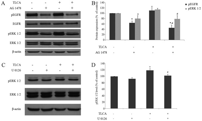
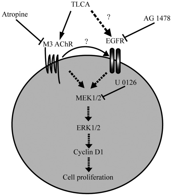
Cholangiocarcinoma (CCA) is a malignant cancer of the biliary tract and its occurrence is associated with chronic cholestasis which causes an elevation of bile acids in the liver and bile duct. The present study aimed to investigate the role and mechanistic effect of bile acids on the CCA cell growth. Intrahepatic CCA cell lines, RMCCA-1 and HuCCA-1, were treated with bile acids and their metabolites to determine the growth promoting effect. Cell viability, cell cycle analysis, EdU incorporation assays were conducted. Intracellular signaling proteins were detected by western immunoblotting. Among eleven forms of bile acids and their metabolites, only taurolithocholic acid (TLCA) concentration dependently (1-40 µM) increased the cell viability of RMCCA-1, but not HuCCA-1 cells. The cell cycle analysis showed induction of cells in the S phase and the EdU incorporation assay revealed induction of DNA synthesis in the TLCA-treated RMCCA-1 cells. Moreover, TLCA increased the phosphorylation of EGFR, ERK 1/2 and also increased the expression of cyclin D1 in RMCCA-1 cells. Furthermore, TLCA-induced RMCCA-1 cell growth could be inhibited by atropine, a non-selective muscarinic acetylcholine receptor (mAChR) antagonist, AG 1478, a specific EGFR inhibitor, or U 0126, a specific MEK 1/2 inhibitor. These results suggest that TLCA induces CCA cell growth via mAChR and EGFR/EKR1/2 signaling pathway. Moreover, the functional presence of cholinergic system plays a certain role in TLCA-induced CCA cell growth.


Figures

Figure 1
Figure 1
Comparison of COX-2 and cholinergic components in HuCCA-1 and RMCCA-1. (A) Western blotting of COX-2 protein and band density compared between HuCCA-1 and RMCCA-1 (*p<0.05 as compared with RMCCA-1). (B) Western blotting of cholinergic component proteins in SH-SY5Y (used as a positive control), RMCCA-1 and HuCCA-1 cells; CHT, choline transporter; ChAT, choline acetyltransferase; AChE, acetylcholine esterase; M3 mAChR, M3 muscarinic acetylcholine receptor; α7 nAChR, α7 nicotinic acetylcholine receptor.
Figure 2
Figure 2
PrestoBlue cell viability assay of RMCCA-1 cells treated with TLCA at 1, 5, 10, 20, 40, 80 and 100 μM for 48 h (*p<0.05 as compared with control)
Figure 3
Figure 3
TLCA effects on the growth of RMCCA-1 cells. RMCCA-1 cells were treated with fetal bovine serum (10% v/v) or TLCA (10 μM) for 24 h and then stained with propidium iodide. Cell cycle was analyzed by ModFit LT software. (A) Cell distribution of cell cycle analysis. (B) RMCCA-1 cell proliferation was determined by using EdU incorporation assay (*p<0.05 as compared with control).
Figure 4
Figure 4
Effect of TLCA on cyclin D1 and pERK 1/2. (A) Western blot bands of cyclin D1, pERK 1/2, ERK 1/2 and β-actin protein of RMCCA-1 cells treated with TLCA for 48 h. (B) The band density ratio of cyclin D1 and pERK 1/2 protein was determined (*p<0.05 as compared with control).
Figure 5
Figure 5
Effects of TLCA on EGFR and ERK. (A) W estern blot bands of pEGFR, EGFR, pERK 1/2, ERK 1/2 and β-actin protein of RMCCA-1 cells treated with TLCA 10 μM at varying times. (B) The band density ratio of pEGFR and pERK 1/2 protein was determined (*p<0.05 as compared with time 0 min).
Figure 6
Figure 6
Effect of acetylcholine receptor agonists and TLCA on CCA cell lines. (A) PrestoBlue cell viability of HuCCA-1 and RMCCA-1 cells treated with carbachol for 48 h. (B) PrestoBlue cell viability of HuCCA-1 and RMCCA-1 cells treated with oxotremorine for 48 h. (C) PrestoBlue cell viability of RMCCA-1 cells treated with TLCA and/or atropine for 48 h (*p<0.05 as compared with control; #p<0.05 as compared with TLCA 10 μM).
Figure 7
Figure 7
M3 AChR did not transactivate the EGFR. (A) Western blot bands of pEGFR, EGFR, pERK 1/2, ERK 1/2 and β-actin protein of RMCCA-1 cells treated with TLCA 10 μM and/or atropine 10 μM at 1 h. (B) The band density ratio of pEGFR and pERK 1/2 protein (*p<0.05 as compared with control).
Figure 8
Figure 8
TLCA effects on EGFR and MAP kinase of RMCCA-1 cells. PrestoBlue cell viability assay was used. (A) RMCCA-1 cells were treated with EGF 100 ng/ml or TLCA 10 μM with or without co-incubation AG 1478 for 24 h. (B) RMCCA-1 cells were treated with TLCA 10 μM and/or U 0126 for 48 h (*p<0.05 was compared with control; #p<0.05 as compared with EGF 100 ng/ml or TLCA 10 μM).
Figure 9
Figure 9
TLCA activates MAP kinase via the EGFR receptor. (A) Western blot bands of pEGFR, EGFR, pERK 1/2, ERK 1/2 and β-actin protein of RMCCA-1 cells treated with TLCA 10 μM and/or AG 1478 0.1 μM at 6 h. (B) The band density ratio of pEGFR and pERK 1/2 protein. (C) Western blot bands of pERK 1/2, ERK1/2, and β-actin protein of RMCCA-1 cells treated with TLCA 10 μM and/or U 0126 0.1 μM at 24 h. (D) The band density ratio of pERK 1/2 protein (*p<0.05 as compared with control; #p<0.05 as compared with TLCA 10 μM).
Figure 10
Figure 10
Proposed diagram of signaling pathways of TLCA mediated RMCCA-1 proliferation. TLCA binds to M3 AChR, which can be blocked by a non-selective mAChR antagonist (atropine). TLCA activates EGFR, which can be blocked by EGFR kinase inhibitor (AG 1478). Activation of M3 AChR and EGFR resulted in activated downstream MAP kinase signaling, which can be blocked by MEK 1/2 inhibitor (U 0126).

Similar articles

Cited by

References

    1. Patel T. Cholangiocarcinoma - controversies and challenges. Nat Rev Gastroenterol Hepatol. 2011;8:189–200. doi: 10.1038/nrgastro.2011.20. - DOI - PMC - PubMed
    1. Raufman JP, Cheng K, Zimniak P. Activation of muscarinic receptor signaling by bile acids: Physiological and medical implications. Dig Dis Sci. 2003;48:1431–1444. doi: 10.1023/A:1024733500950. - DOI - PubMed
    1. Bernstein H, Bernstein C, Payne CM, Dvorakova K, Garewal H. Bile acids as carcinogens in human gastrointestinal cancers. Mutat Res. 2005;589:47–65. doi: 10.1016/j.mrrev.2004.08.001. - DOI - PubMed
    1. Cheng K, Raufman JP. Bile acid-induced proliferation of a human colon cancer cell line is mediated by transactivation of epidermal growth factor receptors. Biochem Pharmacol. 2005;70:1035–1047. doi: 10.1016/j.bcp.2005.07.023. - DOI - PubMed
    1. Fujino T, Takeuchi A, Maruko-Ohtake A, Ohtake Y, Satoh J, Kobayashi T, Tanaka T, Ito H, Sakamaki R, Kashimura R, et al. Critical role of farnesoid X receptor for hepatocellular carcinoma cell proliferation. J Biochem. 2012;152:577–586. doi: 10.1093/jb/mvs101. - DOI - PubMed

Publication types

MeSH terms