. 2015 Mar 27;11(3):e1005120.
doi: 10.1371/journal.pgen.1005120. eCollection 2015 Mar.

Systems biology of tissue-specific response to Anaplasma phagocytophilum reveals differentiated apoptosis in the tick vector Ixodes scapularis

Affiliations

Systems biology of tissue-specific response to Anaplasma phagocytophilum reveals differentiated apoptosis in the tick vector Ixodes scapularis

Nieves Ayllón et al. PLoS Genet. .

Abstract

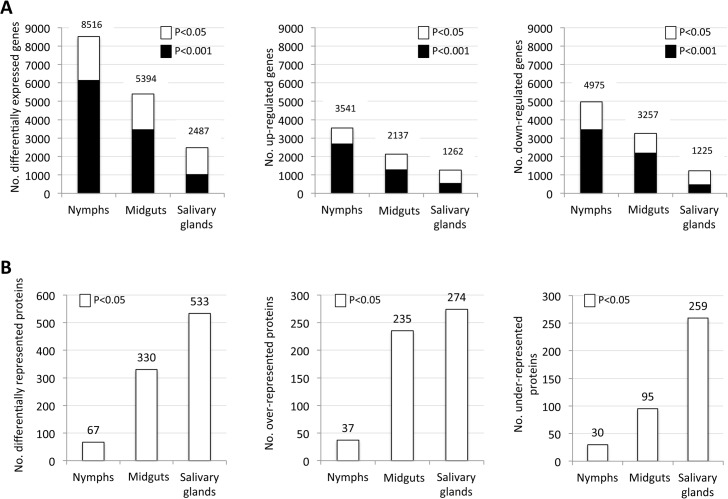
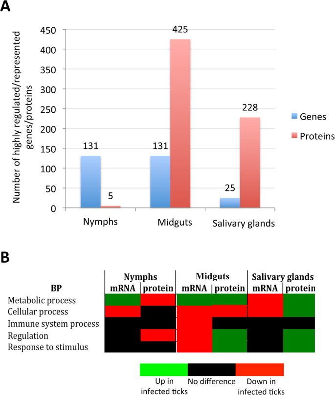
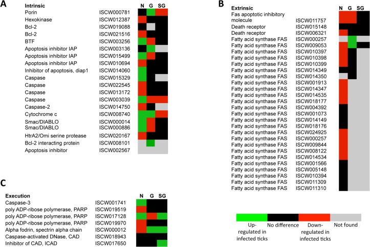
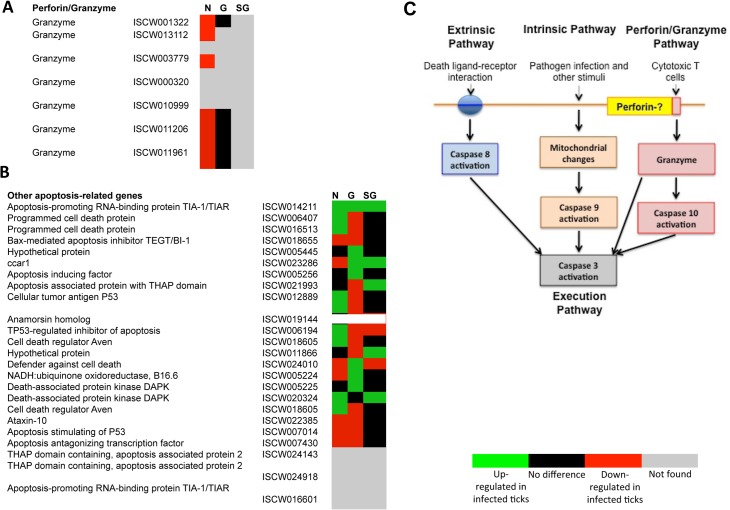
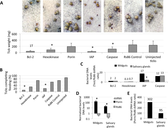
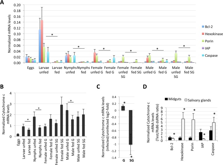
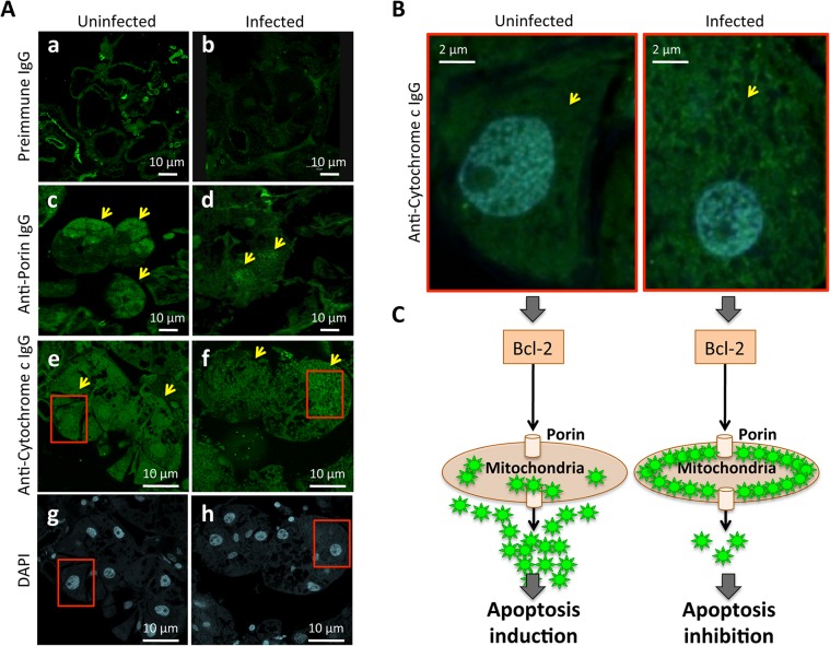
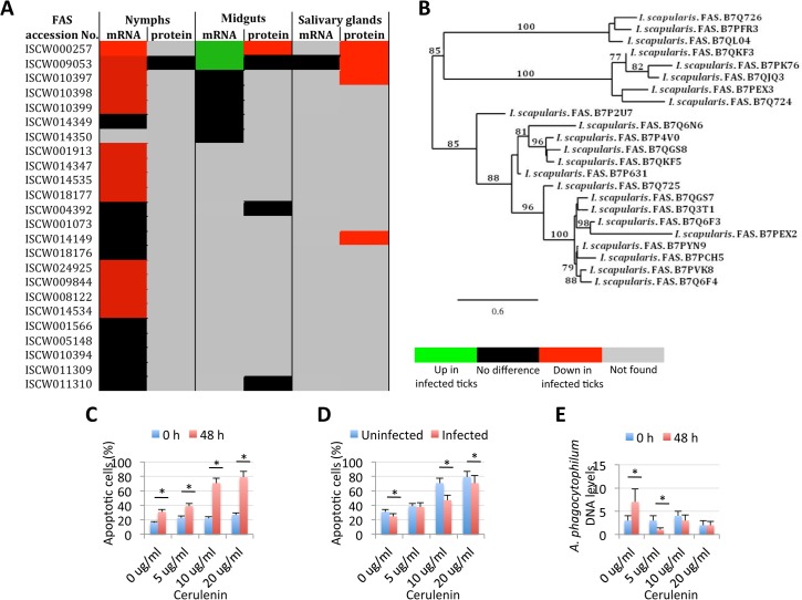
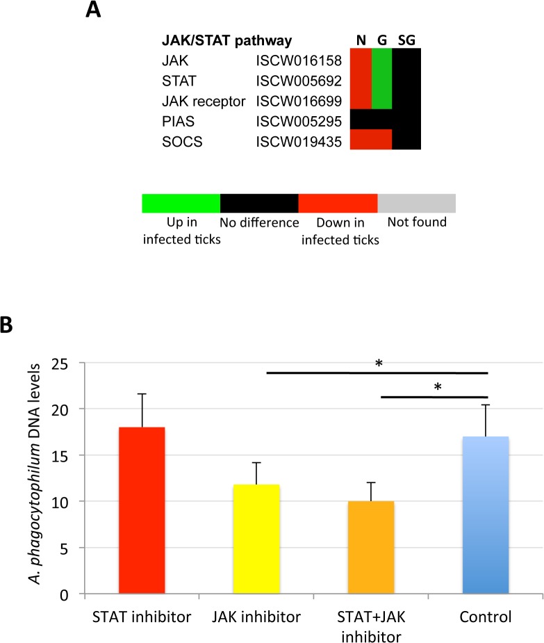
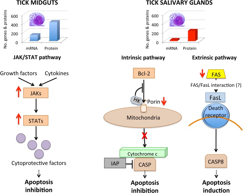
Anaplasma phagocytophilum is an emerging pathogen that causes human granulocytic anaplasmosis. Infection with this zoonotic pathogen affects cell function in both vertebrate host and the tick vector, Ixodes scapularis. Global tissue-specific response and apoptosis signaling pathways were characterized in I. scapularis nymphs and adult female midguts and salivary glands infected with A. phagocytophilum using a systems biology approach combining transcriptomics and proteomics. Apoptosis was selected for pathway-focused analysis due to its role in bacterial infection of tick cells. The results showed tissue-specific differences in tick response to infection and revealed differentiated regulation of apoptosis pathways. The impact of bacterial infection was more pronounced in tick nymphs and midguts than in salivary glands, probably reflecting bacterial developmental cycle. All apoptosis pathways described in other organisms were identified in I. scapularis, except for the absence of the Perforin ortholog. Functional characterization using RNA interference showed that Porin knockdown significantly increases tick colonization by A. phagocytophilum. Infection with A. phagocytophilum produced complex tissue-specific alterations in transcript and protein levels. In tick nymphs, the results suggested a possible effect of bacterial infection on the inhibition of tick immune response. In tick midguts, the results suggested that A. phagocytophilum infection inhibited cell apoptosis to facilitate and establish infection through up-regulation of the JAK/STAT pathway. Bacterial infection inhibited the intrinsic apoptosis pathway in tick salivary glands by down-regulating Porin expression that resulted in the inhibition of Cytochrome c release as the anti-apoptotic mechanism to facilitate bacterial infection. However, tick salivary glands may promote apoptosis to limit bacterial infection through induction of the extrinsic apoptosis pathway. These dynamic changes in response to A. phagocytophilum in I. scapularis tissue-specific transcriptome and proteome demonstrated the complexity of the tick response to infection and will contribute to characterize gene regulation in ticks.


Conflict of interest statement

The authors have declared that no competing interests exist.

Figures

Fig 1
Fig 1. Tissue-specific differentially expressed genes and represented proteins in response to A. phagocytophilum infection.
Tissue-specific differences in tick response to pathogen infection were found at both mRNA and protein levels. (A) Number of differentially expressed genes (total, up-regulated and down-regulated) in infected nymphs, adult female midguts and salivary glands. (B) Number of differentially represented proteins (total, over-represented and under-represented) in infected nymphs, adult female midguts and salivary glands (-2>Zq>2).
Fig 2
Fig 2. Tissue-specific effect of A. phagocytophilum infection on tick biological processes.
The impact of bacterial infection on tick gene expression was more pronounced in nymphs and adult female midguts than in adult female salivary glands, probably reflecting pathogen developmental cycle. (A) Top differentially expressed genes were selected as those with more than 50-fold (log2 normalized fold change > 5.64) difference between infected and uninfected ticks. Top differentially represented proteins were selected as those with more than 5-fold (log2 normalized fold change > 2.32) difference between infected and uninfected ticks. (B) Predominant effect of bacterial infection on tick BPs. The number of top differentially expressed/represented genes/proteins in selected BPs was normalized against the total number of genes/proteins highly expressed/represented in nymphs, adult female midguts and salivary glands to select the predominant effect of bacterial infection on tick BPs.
Fig 3
Fig 3. Annotation of intrinsic, extrinsic and execution apoptosis pathway genes and expression in response to A. phagocytophilum infection.
Tissue-specific differences were found in the apoptosis pathway genes response to A. phagocytophilum infection. (A) Intrinsic pathway. (B) Extrinsic pathway. (C) Execution pathway. For annotation, gene identifiers were obtained from VectorBase (www.vectorbase.org) and compared to the corresponding pathways in Drosophila melanogaster, Anopheles gambiae, Aedes aegypti and Homo sapiens. Differential expression (P<0.05) is shown for tick nymphs (N) and adult female midguts (G) and salivary glands (SG).
Fig 4
Fig 4. Annotation of Perforin/Granzyme and other apoptosis pathway genes and expression in response to A. phagocytophilum infection.
All apoptosis pathways described in other organisms were identified in I. scapularis, except for the absence of the Perforin ortholog in the Perforin/Granzyme pathway. (A) Perforin/Granzyme pathway. (B) Other apoptosis-related genes. For annotation, gene identifiers were obtained from VectorBase (www.vectorbase.org) and compared to the corresponding pathways in Drosophila melanogaster, Anopheles gambiae, Aedes aegypti and Homo sapiens. Differential expression (P<0.05) is shown for tick nymphs (N) and adult midguts (G) and salivary glands (SG). (C) Schematic representation of predicted apoptosis pathways in I. scapularis.
Fig 5
Fig 5. Characterization of apoptosis differentially expressed genes and represented proteins in response to A. phagocytophilum infection in I. scapularis.
Apoptosis pathway gene and protein levels increased or decreased in a tissue-specific manner in response to pathogen infection, thus supporting the functional characterization of this biological pathway in infected in ticks. (A) Differentially expressed apoptosis genes in infected nymphs and adult female midguts and salivary glands. (B) Apoptosis genes/proteins identified by both transcriptomics and proteomics analyses in nymphs and adult female midguts and salivary glands. (C) Comparison between mRNA and proteins levels of selected intrinsic apoptosis pathway components in tick samples in response to pathogen infection. (D) Intrinsic apoptosis pathway genes selected for functional characterization by RNAi. The genes selected for RNAi are shown with the accession number next to them. Abbreviations: HK, Hexokinase; IAP, apoptosis inhibitor; CASP, Caspase.
Fig 6
Fig 6. Intrinsic apoptosis gene knockdown phenotype in ticks injected with dsRNA.
The knockdown of selected intrinsic apoptosis pathway genes had the greatest effects on tick feeding to repletion but, apparently, not on tick attachment. Porin silencing significantly increased tick colonization by A. phagocytophilum but did not affect tick feeding. Methods: Twenty adult female ticks were injected with gene-specific dsRNAs, the unrelated Rs86 dsRNA control or were left uninjected. After dsRNA injection, female ticks were allowed to feed on sheep inoculated intravenously with A. phagocytophilum (NY18 isolate). Female ticks were allowed to feed until full engorgement and tick weight and mortality were determined in individual female ticks collected after feeding. (A) Tick weight was compared between ticks injected with test genes dsRNA and Rs86 control dsRNA by Student's t-test with unequal variance (*P≤0.05). Panels show representative images at day 5 of tick feeding. (B) The number of ticks completing feeding was compared between ticks injected with test genes dsRNA and Rs86 control dsRNA by one-tailed Fisher's exact test (*P≤0.05). (C-E) A. phagocytophilum DNA levels were determined by msp4 real-time PCR normalizing against tick 16S rDNA and shown as test dsRNA to Rs86 dsRNA ratio. Normalized Ct values were compared between ticks injected with test genes dsRNA and Rs86 control dsRNA by Student's t-test with unequal variance (*P<0.05) and were significantly different for Porin dsRNA-injected ticks only. Abbreviations: 1T, only one tick completed feeding; IAP, apoptosis inhibitor.
Fig 7
Fig 7. Expression of selected intrinsic apoptosis pathway genes.
The analysis highlighted a complex mechanism by which tick cells respond to changes in the expression of intrinsic apoptosis pathway genes and suggested an effect of tick feeding on Porin expression that may also contributed to Porin down-regulation in infected adult female salivary glands. (A) Normalized Bcl-2, Hexokinase, Porin, IAP and Caspase mRNA levels (Ct values are shown as Ave+SD) in different tick developmental stages and tissues. (B) Normalized Cytochrome c mRNA levels are shown as Ave+SD in different tick developmental stages and tissues. The mRNA levels were characterized in tick eggs (three batches of approximately 500 eggs each), fed and unfed larvae (three pools of 50 larvae each), fed and unfed nymphs (three pools of 15 nymphs each), and fed and unfed males and females adults tick tissues (4 ticks each) by real-time RT-PCR normalizing against tick cyclophilin and ribosomal protein S4. Porin and Cytochrome c normalized Ct values were compared between fed and unfed ticks by Student's t-test with unequal variance (*P<0.05; N = 3–4). (C) Cytochrome c mRNA levels were determined in infected and uninfected female midguts (G) and salivary glands (SG), normalized against tick tick cyclophilin and ribosomal protein S4, represented as infected/uninfected Log2-fold ratio (Ave+SD) and normalized Ct values compared between infected and uninfected ticks by Student's t-test with unequal variance (*P<0.05; N = 10). (D) Cytochrome c mRNA levels were determined in midguts and salivary glands of ticks with gene knockdown, normalized against tick tick cyclophilin and ribosomal protein S4, represented as test/dsRNA ratio (Ave+SD) and normalized Ct values compared between ticks injected with test genes dsRNA and Rs86 control dsRNA by Student's t-test with unequal variance (*P<0.05). Abbreviation: IAP, apoptosis inhibitor.
Fig 8
Fig 8. Immunohistochemical localization of tick Porin and Cytochrome c.
The results showed that Porin levels were lower in infected than in uninfected tick salivary glands but demonstrated that differences in Cytochrome c levels were not at the protein level but in the localization of Cytochrome c, which was distributed in the cell cytoplasm of uninfected tick salivary glands but was mainly localized within organelles that probably corresponded to mitochondria in A. phagocytophilum-infected tick salivary glands. (A) Representative images of immunofluorescence analysis of uninfected and A. phagocytophilum-infected adult female tick salivary glands. Tick tissues were stained with rabbit anti-tick proteins antibodies (green, FITC). Arrows show positive staining in response to anti-Porin and anti-Cytochrome c IgG not present after incubation with preimmune IgG. (a-b) preimmune control serum-treated cells, which gave similar results between uninfected and infected ticks. (c-d) uninfected and infected cells stained with anti-Porin antibodies. (e-f) uninfected and infected cells stained with anti-Cytochrome c antibodies. (g-h) uninfected and infected cells stained with DAPI. Bars, 10 μm. (B) Sections in the red squares in e-h were magnified 5X and superimposed to illustrate the localization of the Cytochrome c in the cytoplasm of uninfected and infected adult female tick salivary glands. Arrows show positive staining. (C) Model of the Porin-mediated inhibition of Cytochrome c release as the anti-apoptotic mechanism to facilitate bacterial infection of tick salivary glands.
Fig 9
Fig 9. Role of tick Fatty acid synthase (FAS) in response to A. phagocytophilum infection.
The expression of FAS genes was down-regulated in tick salivary glands, suggesting a response to A. phagocytophilum infection by promoting apoptosis to limit bacterial infection through induction of the extrinsic apoptosis pathway. Cerulenin had an effect on cultured tick cells by promoting apoptosis through FAS inhibition and results corroborated the effect of FAS inhibition on reducing A. phagocytophilum infection of tick salivary glands by activating the extrinsic apoptosis pathway in response to bacterial infection. The JAK/STAT pathway genes were down-regulated in nymphs, up-regulated in adult female midguts and not affected by bacterial infection in adult female salivary glands, suggesting that A. phagocytophilum infection decreased immunity in nymphs while inhibited cell apoptosis in midgut cells to facilitate and establish infection. (A) Comparison of FAS mRNA and protein levels in tick samples in response to pathogen infection. (B) Phylogenetic analysis of I. scapularis FAS amino acid sequences. The tree was constructed using the maximum likelihood method. Bootstrap values are represented as percent on internal branches (1000 replicates). Only Bootstrap values higher than 70 are shown. The GenBank accession numbers of the sequences used in the phylogenetic analysis are shown. (C) The percent of apoptotic cells was determined by flow cytometry in uninfected ISE6 tick cells treated for 48 h with different concentrations of the FAS inhibitor, Cerulenin. Results were represented as Ave+SD and compared between cells analyzed at 0 and 48 h of Cerulenin treatment by Student's t-test with unequal variance (*P<0.05; N = 3). (D) The percent of apoptotic cells was determined by flow cytometry in A. phagocytophilum-infected ISE6 tick cells treated for 48 h with different concentrations of the FAS inhibitor, Cerulenin. Results were represented as Ave+SD and compared between uninfected and infected cells by Student's t-test with unequal variance (*P<0.05; N = 3). (E) A. phagocytophilum DNA levels were determined in infected ISE6 tick cells treated for 48 h with different concentrations of the FAS inhibitor, Cerulenin by msp4 real-time PCR normalizing against tick 16S rDNA. Results are shown as Ave+SD normalized Ct values and were compared between cells analyzed at 0 and 48 h of Cerulenin treatment and bacterial infection by Student's t-test with unequal variance (*P<0.05; N = 3).
Fig 10
Fig 10. Role of tick JAK/STAT pathway in response to A. phagocytophilum infection.
The JAK/STAT pathway genes were down-regulated in nymphs, up-regulated in adult female midguts and not affected by bacterial infection in adult female salivary glands, suggesting as corroborated by the treatment of infected tick cells with JAK-STAT inhibitors, that A. phagocytophilum infection decreases immunity in nymphs while inhibiting cell apoptosis in midgut cells to facilitate and establish infection. (A) Annotation of JAK/STAT pathway genes and expression in response to A. phagocytophilum infection. For annotation, gene identifiers were obtained from VectorBase (www.vectorbase.org) and compared to the corresponding pathways in Drosophila melanogaster, Anopheles gambiae, Aedes aegypti and Homo sapiens. Differential expression (P<0.05) is shown for tick nymphs (N) and adult midguts (G) and salivary glands (SG). (B) A. phagocytophilum DNA levels were determined in infected ISE6 tick cells untreated (Control) or treated for 48 h with STAT (9.2 μM), JAK (400 nM), and STAT (9.2 μM) + JAK (400 nM) inhibitors. Bacterial DNA levels were determined by msp4 real-time PCR normalizing against tick 16S rDNA. Results are shown as Ave+SD normalized Ct values and were compared between untreated and treated cells by Student's t-test with unequal variance (*P<0.05; N = 4).
Fig 11
Fig 11. Effect of A. phagocytophilum infection on adult female midguts and salivary glands.
The number of highly differentially expressed genes and represented proteins was higher in tick midguts than in salivary glands, suggesting a higher impact of bacterial infection in midgut cells probably associated with A. phagocytophilum developmental cycle. In tick midguts, A. phagocytophilum inhibited cell apoptosis to facilitate and establish infection through up-regulation of the JAK/STAT pathway genes. In tick salivary glands, down-regulation of Porin resulted in the inhibition of the Cytochrome c release that inhibited the mitochondrially-induced intrinsic apoptosis pathway to facilitate bacterial infection. This effect was contrasted in part by the induction of the extrinsic apoptosis pathway through the inhibition of FAS proteins.

References

    1. de la Fuente J, Estrada-Peña A, Venzal JM, Kocan KM, Sonenshine DE (2008) Overview: Ticks as vectors of pathogens that cause disease in humans and animals. Front Biosci 13: 6938–6946. - PubMed
    1. Reichard MV, Manzano-Roman R, Kocan KM, Blouin EF, de la Fuente J, et al. (2009) Inoculation of white-tailed deer (Odocoileus virginianus) with Ap-V1 or NY-18 strains of Anaplasma phagocytophilum and microscopic demonstration of Ap-V1 in Ixodes scapularis adults that acquired infection from deer as nymphs. Vector Borne Zoonotic Dis 9: 565–568. 10.1089/vbz.2008.0106 - DOI - PubMed
    1. Sukumaran B, Narasimham S, Anderson JF, DePonte K, Marcantonio K, et al. (2006) An Ixodes scapularis protein required for survival of Anaplasma phagocytophilum in tick salivary glands. J Exp Med 6: 1507–1517. - PMC - PubMed
    1. Dumler JS, Barbet AC, Bekker CPJ, Dasch GA, Palmer GH, et al. (2001) Reorganization of the genera in the families Rickettsiaceae and Anaplasmataceae in the order Rickettsiales: unification of some species of Ehrlichia with Anaplasma, Cowdria with Ehrlichia and Ehrlichia with Neorickettsia, descriptions subjective synonyms of Ehrlichia phagocytophila . Int J Syst Evol Microbiol 51: 2145–2165. - PubMed
    1. Rikihisa Y (2011) Molecular pathogensis of Anaplasma phagocytophilum . Current Microbiol Rev 24: 469–489. 10.1128/CMR.00064-10 - DOI - PMC - PubMed

Publication types

MeSH terms

LinkOut - more resources