. 2015 Mar 27;10(3):e0120893.
doi: 10.1371/journal.pone.0120893. eCollection 2015.

Hypothermia augments neuroprotective activity of mesenchymal stem cells for neonatal hypoxic-ischemic encephalopathy

Affiliations

Hypothermia augments neuroprotective activity of mesenchymal stem cells for neonatal hypoxic-ischemic encephalopathy

Won Soon Park et al. PLoS One. .

Abstract

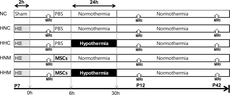
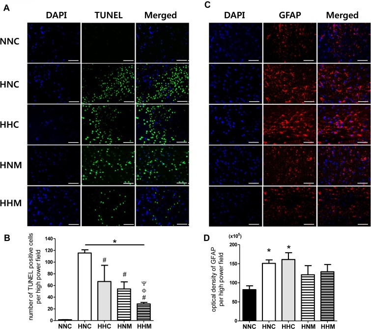
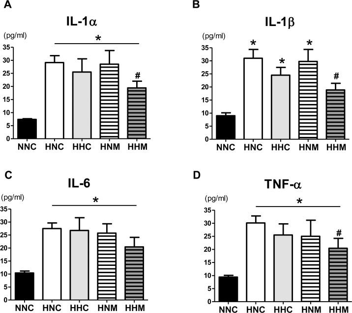
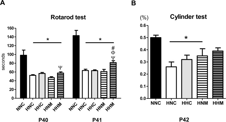
Though hypothermia is the only clinically available treatment for neonatal hypoxic-ischemic encephalopathy (HIE), it is not completely effective in severe cases. We hypothesized that combined treatment with hypothermia and transplantation of human umbilical cord blood (UCB)-derived mesenchymal stem cells (MSCs) would synergistically attenuate severe HIE compared to stand-alone therapy. To induce hypoxia-ischemia (HI), male Sprague-Dawley rats were subjected to 8% oxygen for 120 min after unilateral carotid artery ligation on postnatal day (P) 7. After confirmation of severe HIE involving >50% of the ipsilateral hemisphere volume as determined by diffusion-weighted brain magnetic resonance imaging (MRI) within 2 h after HI, intraventricular MSC transplantation (1 × 10⁵ cells) and/or hypothermia with target temperature at 32°C for 24 h were administered 6 h after induction of HI. Follow-up brain MRI at P12 and P42, sensorimotor function tests at P40-42, evaluation of cytokines in the cerebrospinal fluid (CSF) at P42, and histologic analysis of peri-infarct tissues at P42 were performed. Severe HI resulted in progressively increased brain infarction over time as assessed by serial MRI, increased number of cells positive for terminal deoxynucleotidyl transferase nick-end labeling, microgliosis and astrocytosis, increased CSF cytokine levels, and impaired function in behavioral tests such as rotarod and cylinder tests. All of the abnormalities observed in severe HIE showed greater improvement after combined treatment with hypothermia and MSC transplantation than with either therapy alone. Overall, these findings suggest that combined treatment with hypothermia and human UCB-derived MSC transplantation might be a novel therapeutic modality to improve the prognosis of severe HIE, an intractable disease that currently has no effective treatment.


Conflict of interest statement

Competing Interests: Soo Jin Choi declares the following potential conflict of interest situation arising from the paid employment from the Medipost, Ltd. This does not alter the authors' adherence to PLOS ONE policies on sharing data and materials.

Figures

Fig 1
Fig 1. Experimental protocol.
Fig 2
Fig 2. Combined hypothermia and mesenchymal stem cell transplantation attenuated the progression of ischemic brain lesions after severe HIE.
(A) Representative serial brain MRI images from treatment groups at P7, P12, and P42 (0, 5, and 35 days after inducing HIE, respectively). (B) Volume ratio of the ipsilateral intact area to the contralateral contralateral hemisphere area measured by MRI. Data are mean ± SEM. HNC, HIE+normothermia control group (n = 8); HHC, HIE+hypothermia group (n = 7); HNM, HIE+normothermia+MSCs group (n = 9); HHM, HIE+hypothermia+MSCs group (n = 8). # P < 0.05 vs. HNC.
Fig 3
Fig 3. Combined hypothermia and mesenchymal stem cell transplantation ameliorated the increase in cell death and reactive gliosis in the brain after severe HIE.
The images show representative immunofluorescence micrographs of the penumbra area with staining for TUNEL (A), glial fibrillary acidic protein (GFAP) (C), and DAPI (original magnification; ×400, scale bars; 25 μm). Data are average number of TUNEL-positive cells (B) and average density of GFAP staining (D) in the penumbra area, presented as mean ± SEM. NNC, sham control group (n = 5); HNC, HIE+normothermia control group (n = 8); HHC, HIE+hypothermia group (n = 7); HNM, HIE+normothermia+MSCs group (n = 9); HHM, HIE+hypothermia+MSCs group (n = 8). * P < 0.05 vs. NNC, # P < 0.05 vs. HNC, Φ P < 0.05 vs. HHC, Ψ P < 0.05 vs. HNM.
Fig 4
Fig 4. Combined hypothermia and mesenchymal stem cell transplantation reduced brain inflammation as represented by active macrophages after severe HIE.
(A) Representative immunofluorescence micrographs of the penumbra area with staining for ED-1 (red) and DAPI (blue) (original magnification; ×400, scale bars; 25 μm). Average optical density of ED-1 staining (B) in the penumbra area. Data are mean ± SEM. NNC, sham control group (n = 5); HNC, HIE+normothermia control group (n = 8); HHC, HIE+hypothermia group (n = 7); HNM, HIE+normothermia+MSCs group (n = 9); HHM, HIE+hypothermia+MSCs group (n = 8). *P < 0.05 vs. NNC, # P < 0.05 vs. HNC.
Fig 5
Fig 5. Concentrations of inflammatory cytokines IL-1α, IL-β, IL-6, and TNF- α in cerebrospinal fluid at P42.
Data are mean ± SEM. NNC, sham control group (n = 5); HNC, HIE+normothermia control group (n = 8); HHC, HIE+hypothermia group (n = 7); HNM, HIE+normothermia+MSCs group (n = 9); HHM, HIE+hypothermia+MSCs group (n = 8). *P < 0.05 vs. NNC, # P < 0.05 vs. HNC.
Fig 6
Fig 6. Combined hypothermia and mesenchymal stem cells transplantation improved impaired sensorimotor function after severe HIE.
Sensorimotor functional outcomes on rotarod (A) and cylinder (B) tests. Data are mean ± SEM. NNC, sham control group (n = 5); HNC, HIE+normothermia control group (n = 8); HHC, HIE+hypothermia group (n = 7); HNM, HIE+normothermia+MSCs group (n = 9); HHM, HIE+hypothermia+MSCs group (n = 8). * P< 0.05 vs. NNC, # P < 0.05 vs. HNC, Φ < 0.05 vs. HHC, Ψ P < 0.05 vs. HNM.

Similar articles

Cited by

References

    1. Johnston MV. Hypoxic and ischemic disorders of infants and children. Lecture for 38th meeting of Japanese Society of Child Neurology, Tokyo, Japan, July 1996. Brain Dev. 1997;19: 235–239. - PubMed
    1. Robertson C, Finer N. Term infants with hypoxic-ischemic encephalopathy: outcome at 3.5 years. Dev Med Child Neurol. 1985;27: 473–484. - PubMed
    1. Yenari M, Kitagawa K, Lyden P, Perez-Pinzon M. Metabolic downregulation: a key to successful neuroprotection? Stroke. 2008;39: 2910–2917. 10.1161/STROKEAHA.108.514471 - DOI - PMC - PubMed
    1. Jacobs S, Hunt R, Tarnow-Mordi W, Inder T, Davis P. Cooling for newborns with hypoxic ischaemic encephalopathy. Cochrane Database Syst Rev. 2007: CD003311. - PubMed
    1. Shankaran S, Laptook A, Wright LL, Ehrenkranz RA, Donovan EF, Fanaroff AA, et al. Whole-Body Hypothermia for Neonatal Encephalopathy: Animal Observations as a Basis for a Randomized, Controlled Pilot Study in Term Infants. Pediatrics. 2002;110: 377–385. - PubMed

Publication types

Substances