. 2015 Mar 27;10(3):e0121491.
doi: 10.1371/journal.pone.0121491. eCollection 2015.

A GFP expressing influenza A virus to report in vivo tropism and protection by a matrix protein 2 ectodomain-specific monoclonal antibody

Affiliations

A GFP expressing influenza A virus to report in vivo tropism and protection by a matrix protein 2 ectodomain-specific monoclonal antibody

Sarah De Baets et al. PLoS One. .

Abstract

The severity of influenza-related illness is mediated by many factors, including in vivo cell tropism, timing and magnitude of the immune response, and presence of pre-existing immunity. A direct way to study cell tropism and virus spread in vivo is with an influenza virus expressing a reporter gene. However, reporter gene-expressing influenza viruses are often attenuated in vivo and may be genetically unstable. Here, we describe the generation of an influenza A virus expressing GFP from a tri-cistronic NS segment. To reduce the size of this engineered gene segment, we used a truncated NS1 protein of 73 amino acids combined with a heterologous dimerization domain to increase protein stability. GFP and nuclear export protein coding information were fused in frame with the truncated NS1 open reading frame and separated from each other by 2A self-processing sites. The resulting PR8-NS1(1-73)GFP virus was successfully rescued and replicated as efficiently as the parental PR8 virus in vitro and was slightly attenuated in vivo. Flow cytometry-based monitoring of cells isolated from PR8-NS1(1-73)GFP virus infected BALB/c mice revealed that GFP expression peaked on day two in all cell types tested. In particular respiratory epithelial cells and myeloid cells known to be involved in antigen presentation, including dendritic cells (CD11c+) and inflammatory monocytes (CD11b+ GR1+), became GFP positive following infection. Prophylactic treatment with anti-M2e monoclonal antibody or oseltamivir reduced GFP expression in all cell types studied, demonstrating the usefulness of this reporter virus to analyze the efficacy of antiviral treatments in vivo. Finally, deep sequencing analysis, serial in vitro passages and ex vivo analysis of PR8-NS1(1-73)GFP virus, indicate that this virus is genetically and phenotypically stable.


Conflict of interest statement

Competing Interests: The authors have declared that no competing interests exist.

Figures

Fig 1
Fig 1. In vitro characterization of the PR8-NS1(1–73)GFP virus.
(A) Schematic representation of the promoters and coding sequences of the pHW-NS1(1–73)Dmd-GFP-NEP plasmid used to generate the reporter GFP influenza virus. (B) Multi-cycle growth kinetics. MDCK cells were infected in duplicate with a MOI of 0.001 of wild type PR8 virus or PR8-NS1(1–73)GFP virus in the presence of TPCK-trypsin. At the indicated time points after infection, the viral titer in the supernatant (50 μl sample) was determined by TCID50 analysis. The graph shows the mean with the standard error of each data point. For the wild type PR8 virus, one of the duplicate samples of the 48 h time point was excluded due to technical failure. (C) Plaques of PR8 and PR8-NS1(1–73)GFP virus were visualized on day three after infection of MDCK cells by immunostaining with an M2e-specific monoclonal antibody. (D) Confocal microscopy analysis of MDCK cells infected with wild type PR8 or PR8-NS1(1–73)GFP virus (MOI 1). Twenty four hours after infection the cells were fixed and stained with anti-RNP (red; middle panel) and Hoechst (blue). The GFP signal is shown in green (top panel). An overlay of the three colors is shown in the bottom panel. (E) MDCK cells were infected with a MOI of 1 of PR8-NS1(1–73)GFP virus or wild type PR8 virus, or were not infected (NI). After 24 h, lysates were prepared and the proteins were visualized by western blotting and immune-detection with an anti-GFP (top), anti-HA (middle) or anti-NS1 (bottom) antibody. The panel of anti-HA is split in two parts, as different exposure times were used to reveal the protein bands.
Fig 2
Fig 2. PR8-NS1(1–73)GFP virus is pathogenic in mice.
(A) BALB/c mice (n = 6 per group) were inoculated with 1 x 10³, 1 x 10⁴ or 1 x 10⁵ PFU of PR8-NS1(1–73)GFP virus or 1 x 10³ PFU of wild type PR8 virus. Body weight (relative to initial body weight on day 0) and survival were monitored for 14 days. Error bars represent the standard deviation. The body weight of mice infected with 1 x 10³ or 1 x 10⁵ PFU of PR8-NS1(1–73)GFP virus was significantly different on day 3–7 or day 2–6, respectively, from mice infected with 1 x 10³ PFU of wild type PR8 virus (one-way ANOVA with a Tukey’s multiple comparison test, * P < 0.05, ** P < 0.01, *** P < 0.001). The survival curves of mice infected with 1 x 10³ or 1 x 10⁵ PFU of PR8-NS1(-173)GFP virus were significantly different from mice infected with 1 x 10³ PFU of wild type PR8 virus (log-rank test, * P < 0.05, ** P < 0.01) (B) BALB/c mice (n = 8 per group) were inoculated with 1 x 10³ or 1 x 10⁴ PFU of PR8-NS1(1–73)GFP virus or 1 x 10³ PFU of wild type PR8 virus. Body weight (relative to initial body weight on day 0) was monitored for 5 days. Data points represent averages and error bars represent the standard deviation. Body weight of mice infected with PR8-NS1(1–73)GFP virus was significantly different from those infected with wild type PR8 virus (two-way ANOVA with a Tukey’s multiple comparison test, *** P < 0.001). The virus titer in the lungs was assessed on day 2 and day 5 by TCID50 analysis of the lung homogenates (50 μl sample). For the titers on day two in the wild type PR8 infected group, one value was excluded due to technical failure. Differences in viral titers on day 2 or day 5 were not statistically significant (P > 0.05, one-way ANOVA, with a Tukey’s multiple comparison test).
Fig 3
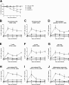
Fig 3. Kinetics and in vivo cell tropism of the PR8-NS1(1–73)GFP virus in mice.
BALB/c mice (n = 19 per group) were infected with 1 x 10⁴ PFU of PR8-NS1(1–73)GFP or 1 x 10³ PFU of wild type PR8 virus. Each day, three mice from each group were euthanized. The lungs were isolated and the number of GFP positive cells was determined using multicolor flow cytometry. (A) Body weight was monitored for five days. Average body weight relative to initial weight on day 0 (n = 7, except for wild type n = 6). Body weights of mice treated with anti-M2e or oseltamivir were significantly different from those of mice treated with anti-NBe (** P < 0.01, *** P < 0.001); two-way ANOVA with a Tukey’s multiple comparison test. (B-J) GFP expression was analyzed in non-immune (CD45-) cells (B), and in immune (CD45+) cells (C), including macrophages (D), T cells (E), B cells (F), NK cells (G), dendritic cells (H-I) and inflammatory monocytes (J). The error bars represent the standard deviation. This graph is representative of two independent experiments.
Fig 4
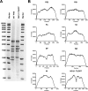
Fig 4. Nucleotide sequence analysis of PR8-NS1(1–73)GFP virus.
(A) Electrophoretic analysis of RT-PCR products generated from viral RNA extracted from virus particles of PR8 (lane 2) and PR8-NS1(1–73)GFP (lane 3). The asterisk indicates the RT-PCR product derived from wild type NS or the engineered NS1(1–73)GFP gene segment. (B) Sequence coverage for the different genome segments of the PR8-NS1(1–73)GFP virus stock as determined by Illumina MiSeq sequencing and CLC genomics version 7.0.3 workbench data processing. The obtained sequences were mapped to the reference genome based on the plasmids used to generate the virus.

References

    1. Everitt AR, Clare S, Pertel T, John SP, Wash RS, Smith SE, et al. IFITM3 restricts the morbidity and mortality associated with influenza. Nature. 2012;484(7395):519–23. 10.1038/nature10921 - DOI - PMC - PubMed
    1. Katz JM, Plowden J, Renshaw-Hoelscher M, Lu X, Tumpey TM, Sambhara S. Immunity to influenza: the challenges of protecting an aging population. Immunologic research. 2004;29(1–3):113–24. - PubMed
    1. Poehling KA, Edwards KM, Griffin MR, Szilagyi PG, Staat MA, Iwane MK, et al. The burden of influenza in young children, 2004–2009. Pediatrics. 2013;131(2):207–16. 10.1542/peds.2012-1255 - DOI - PMC - PubMed
    1. Peiris JS, Hui KP, Yen HL. Host response to influenza virus: protection versus immunopathology. Curr Opin Immunol. 2010;22(4):475–81. 10.1016/j.coi.2010.06.003 - DOI - PMC - PubMed
    1. Sridhar S, Begom S, Bermingham A, Hoschler K, Adamson W, Carman W, et al. Cellular immune correlates of protection against symptomatic pandemic influenza. Nature medicine. 2013;19(10):1305–12. 10.1038/nm.3350 - DOI - PubMed

Publication types

MeSH terms