. 2015 Mar 27;10(3):e0120441.
doi: 10.1371/journal.pone.0120441. eCollection 2015.

Pulmonary inflammation is regulated by the levels of the vesicular acetylcholine transporter

Affiliations

Pulmonary inflammation is regulated by the levels of the vesicular acetylcholine transporter

Nathalia M Pinheiro et al. PLoS One. .

Abstract

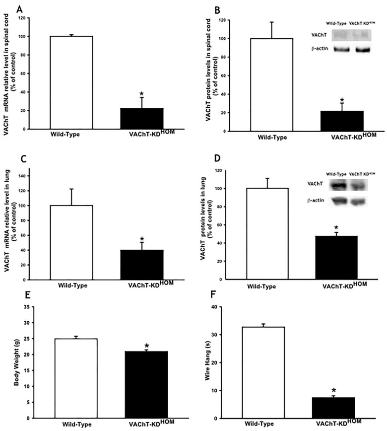
Acetylcholine (ACh) plays a crucial role in physiological responses of both the central and the peripheral nervous system. Moreover, ACh was described as an anti-inflammatory mediator involved in the suppression of exacerbated innate response and cytokine release in various organs. However, the specific contributions of endogenous release ACh for inflammatory responses in the lung are not well understood. To address this question we have used mice with reduced levels of the vesicular acetylcholine transporter (VAChT), a protein required for ACh storage in secretory vesicles. VAChT deficiency induced airway inflammation with enhanced TNF-α and IL-4 content, but not IL-6, IL-13 and IL-10 quantified by ELISA. Mice with decreased levels of VAChT presented increased collagen and elastic fibers deposition in airway walls which was consistent with an increase in inflammatory cells positive to MMP-9 and TIMP-1 in the lung. In vivo lung function evaluation showed airway hyperresponsiveness to methacholine in mutant mice. The expression of nuclear factor-kappa B (p65-NF-kB) in lung of VAChT-deficient mice were higher than in wild-type mice, whereas a decreased expression of janus-kinase 2 (JAK2) was observed in the lung of mutant animals. Our findings show the first evidence that cholinergic deficiency impaired lung function and produce local inflammation. Our data supports the notion that cholinergic system modulates airway inflammation by modulation of JAK2 and NF-kB pathway. We proposed that intact cholinergic pathway is necessary to maintain the lung homeostasis.


Conflict of interest statement

Competing Interests: The authors have declared that no competing interests exist.

Figures

Fig 1
Fig 1. Protein content and mRNA levels of VAChT. A and B.
VAChT mRNA expression (A) measured by real-time PCR (6–8 mice per group) and protein content (B) quantified by Western Blot (3 mice per group) in spinal cord from wild-type and VAChT-KDHOM (mutant). C and D. VAChT mRNA expression (C) measured by real-time PCR (6–8 per group) and protein content (D) quantified by Western blot (6–8 mice per group) in lung from wild-type and VAChT-KDHOM (mutant). Glyceraldehyde 3-phosphate dehydrogenase (GAPDH) was used as a housekeeping for gene expression analysis (A and C) and β-actin was used as protein loading control for Western Blot (B and D). Both were presented as a percentage of WT. The gels (B and D) is representative of results that were obtained in an experiment that was repeated two times. *p<0.05 vs wild-type mice. E and F: Body weight in g (E) in wild-type and mutant mice and the time in wire hang test performed before all measurements in seconds (F). E and F represent data of 8–14 animals per group. Data area expressed as means ± SEM. *P<0.001 vs. wild-type mice.
Fig 2
Fig 2. VAChT deficiency did not affect other cholinergic component in lung.
Muscarinic receptor 2 (M2), high-affinity choline transporter (CHT1), α7 nicotinic acetylcholine receptor (α7nAChR) and Acetylcholinesterase protein expression was analyzed by Western Blot. The gel is representative of results that were obtained in an experiment that was repeated two times. The graphs represent the values normalized by β-actin.
Fig 3
Fig 3. VAChT deficiency increased peribronchial edema and pulmonary inflammation.
A. Peribronchial edema evaluated around airways. Lung were fixed in 10% formalin and embedded in paraffin before sections were cut and stained with hematoxylyn and eosin. VAChT KD-HOM increased peribronchovascular edema (*p<0.001 vs wild-type mice). B to D. Representative photomicrographs illustrating the peribronchial edema and cellular infiltration around airways obtained from a VAChT mutant mice (C and D) compared to wild-type (B). E. Amount of total protein measured in bronchoalveolar lavage (BALF) (n = 7–8 per group, *p<0.01 vs wild-type mice). F. Mean and standard error of macrophages, lymphocytes, eosinophils and neutrophils counted in bronchial alveolar lavage fluid (BALF) (n = 7–8 per group, *P<0.05 vs wild-type mice). G and H represent peribronchovascular mononuclear (*p<0.05 vs wild-type mice) and polymorphonuclear cells, respectively, evaluated around airways.
Fig 4
Fig 4. VAChT-deficient mice presented high levels of pro-inflammatory cytokines.
Data are expressed as mean ±SEM of five to eight mice per group. Cytokines was measured by ELISA in lung homogenate. Mutant mice (VAChT KD-HOM) presented high values of TNF-α and IL-4 compared to wild-type animals. *p<0.05 vs wild-type mice.
Fig 5
Fig 5. VAChT deficiency increased p65-NF-kB expression in lung.
Subunit p65-NF-kB protein expression was measured by western blot (A). The gel is representative of results that were obtained in an experiment that was repeated two times. The graphs represent the values normalized by β-actin (n = 5 per group). *p<0.05 vs wild-type group. Number of inflammatory cells positive to p65-NF-kB (B) from 6–8 animals per group was visualized by immunohistochemistry in paraffin embedded section. Representative photomicrographs used to detect NF-kB (Panels C to F) showed a stronger stain in mutant mice (E and F) compared to wild-type mice (C and D). Arrows indicate positive cells around airway wall. *p<0.001 vs wild-type group.
Fig 6
Fig 6. VAChT deficiency reduced JAK-2 expression in lung.
Janus kinase 2 (JAK-2) (A), signal transducer and activator of transcription 3 (STAT3) (B) and phosphorylated STAT3 (C) and suppressor of cytokine signaling 3 (SOCS-3) (D) protein expression was measured by Western Blot. The gel is representative of results that were obtained in an experiment that was repeated two times. The graphs represent the values normalized by β-actin. *p<0.05 vs wild-type group.
Fig 7
Fig 7. VAChT-deficient induced airway remodeling.
Data of collagen (A) and elastic fibers content (B) are expressed as mean ±SEM at twelve to fourteen mice per group. It was evaluated in paraffin sections stained with Picro-Sirius and Resorcin-Fuchsin respectively, and it was measured around airways using an image analysis system. Number of inflammatory cells positive to MMP-9 (C) and TIMP-1 (D) from 6–8 animals per group was visualized by immunohistochemistry in paraffin embedded section. Collagen content was enhanced by VAChT deficiency and it can be observed in panels E (wild-type) and H (mutant mice). Representative photomicrographs used to detect MMP-9 (Panels F and I) and TIMP-1 (panels G and J) showed a stronger stain in mutant mice. A and D *p<0.001 vs wild-type group and B and C *p<0.05 vs wild-type group.
Fig 8
Fig 8. Airway hyperresponsiveness in VAChT deficiency animals.
Respiratory system elastance (Ers) (A and B) and resistance (Rrs) (C and D) was recorded in wild-type and mutant mice. We performed a dose response curve to methacholine and values were obtained 30 seconds after each infusion (panels B and D). We also analyzed the percentage of maximal responses related to baseline (A and C). Data are expressed as mean and standard error of the 6–8 animals per group. *p<0.05 compared to wild-type.

References

    1. Moldoveanu B, Otmishi P, Jani P, Walker J, Sarmiento X, Guardiola J, et al. Inflammatory mechanisms in the lung. J Inflamm Res. 2009;2: 1–11. - PMC - PubMed
    1. Postma DS, Timens W. Remodeling in asthma and chronic obstructive pulmonary disease. Proc Am Thorac Soc. 2006;3: 434–439. - PubMed
    1. Rosas-Ballina M, Olofsson PS, Ochani M, Valdes-Ferrer SI, Levine YA, Reardon C, et al. Acetylcholine-synthesizing T cells relay neural signals in a vagus nerve circuit. Science. 2011;334: 98–101. 10.1126/science.1209985 - DOI - PMC - PubMed
    1. Su X, Matthay MA, Malik AB. Requisite role of the cholinergic alpha7 nicotinic acetylcholine receptor pathway in suppressing Gram-negative sepsis-induced acute lung inflammatory injury. J Immunol. 2010;184: 401–410. 10.4049/jimmunol.0901808 - DOI - PMC - PubMed
    1. Su X, Lee JW, Matthay ZA, Mednick G, Uchida T, Fang X, et al. Activation of the alpha7 nAChR reduces acid-induced acute lung injury in mice and rats. Am J Respir Cell Mol Biol. 2007;37: 186–192. - PMC - PubMed

Publication types

MeSH terms