Clinical Trial
. 2015 Mar 27;11(3):e1004746.
doi: 10.1371/journal.ppat.1004746. eCollection 2015 Mar.

Early virological and immunological events in asymptomatic Epstein-Barr virus infection in African children

Affiliations
Clinical Trial

Early virological and immunological events in asymptomatic Epstein-Barr virus infection in African children

Shamanthi Jayasooriya et al. PLoS Pathog. .

Abstract

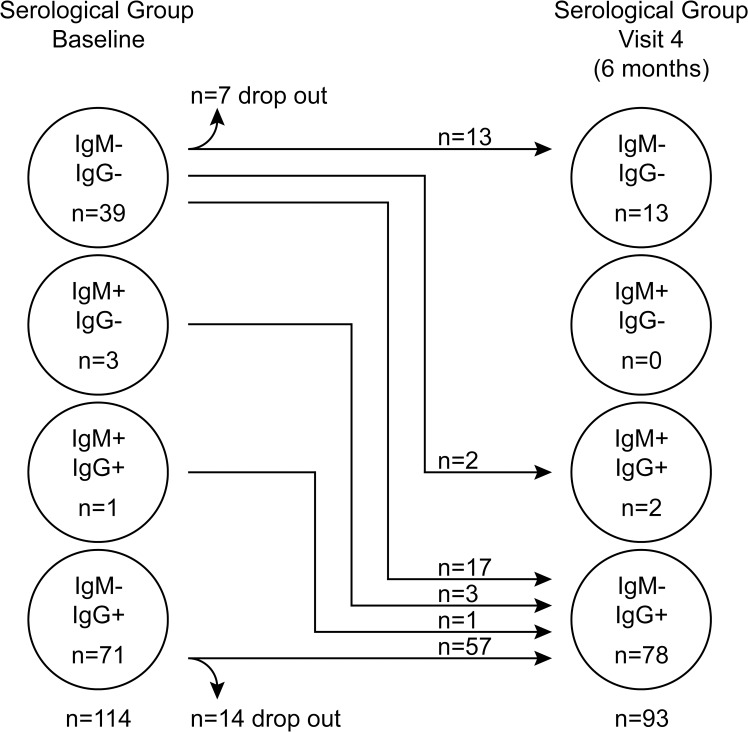
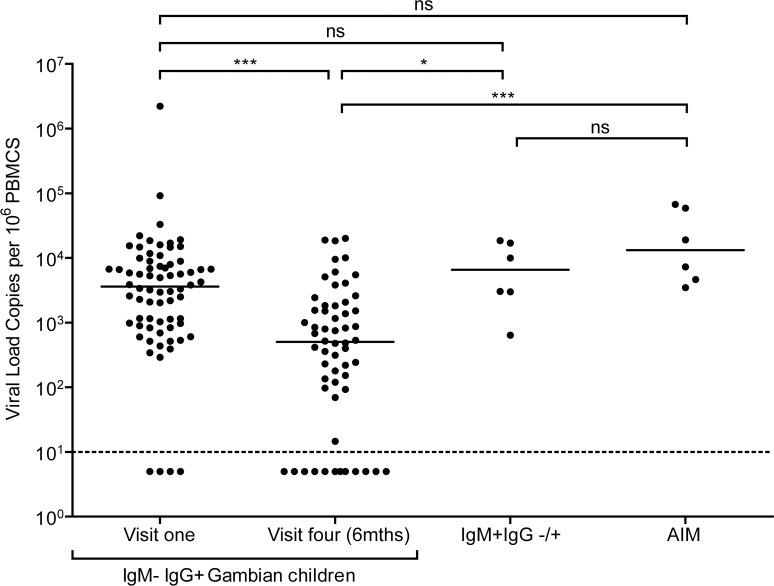
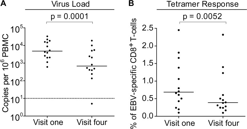
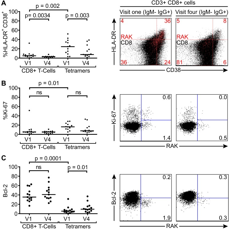
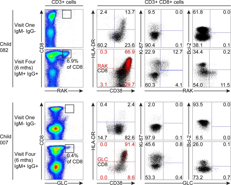
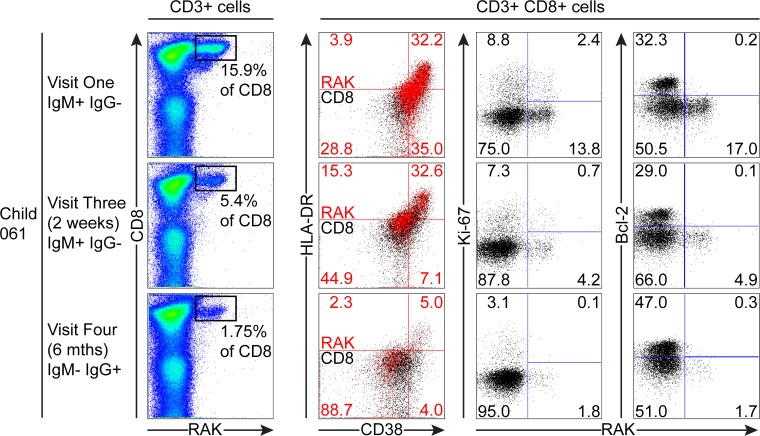
Epstein-Barr virus (EBV) infection often occurs in early childhood and is asymptomatic. However, if delayed until adolescence, primary infection may manifest as acute infectious mononucleosis (AIM), a febrile illness characterised by global CD8+ T-cell lymphocytosis, much of it reflecting a huge expansion of activated EBV-specific CD8+ T-cells. While the events of AIM have been intensely studied, little is known about how these relate to asymptomatic primary infection. Here Gambian children (14-18 months old, an age at which many acquire the virus) were followed for the ensuing six months, monitoring circulating EBV loads, antibody status against virus capsid antigen (VCA) and both total and virus-specific CD8+ T-cell numbers. Many children were IgG anti-VCA-positive and, though no longer IgM-positive, still retained high virus loads comparable to AIM patients and had detectable EBV-specific T-cells, some still expressing activation markers. Virus loads and the frequency/activation status of specific T-cells decreased over time, consistent with resolution of a relatively recent primary infection. Six children with similarly high EBV loads were IgM anti-VCA-positive, indicating very recent infection. In three of these donors with HLA types allowing MHC-tetramer analysis, highly activated EBV-specific T-cells were detectable in the blood with one individual epitope response reaching 15% of all CD8+ T-cells. That response was culled and the cells lost activation markers over time, just as seen in AIM. However, unlike AIM, these events occurred without marked expansion of total CD8+ numbers. Thus asymptomatic EBV infection in children elicits a virus-specific CD8+ T-cell response that can control the infection without over-expansion; conversely, in AIM it appears the CD8 over-expansion, rather than virus load per se, is the cause of disease symptoms.


Conflict of interest statement

The authors have declared that no competing interests exist.

Figures

Fig 1
Fig 1. EBV VCA serological status of Gambian children.
Serological status of study participants at baseline (visit one) and six months later at visit four.
Fig 2
Fig 2. EBV loads in peripheral blood mononuclear cells (PBMCs) from Gambian infants compared to Acute Infectious Mononuclear (AIM) patients.
EBV genome loads in IgM-IgG+ Gambian children at visit one (n = 70) and after six months at visit four (n = 58), compared to IgM+IgG+/- children from either time point. For comparison, data of virus loads measured in Caucasian adolescent AIM patients are also presented. The dashed line represents the lower limit of detection for EBV genomes in the assay. Donors below the dashed line had undetectable viral loads. P values calculated using Dunn’s Multiple Comparison Test (one way analysis of variance (ANOVA)). * p<0.05 ** P<0.01 *** P<0.001
Fig 3
Fig 3. Size of lymphocyte populations in the blood of Gambian children and AIM patients.
Absolute numbers of selected T and B-cell subsets were measured from EBV non-infected (IgM-IgG-), established infection (IgM-IgG+) or recently infected (IgM+IgG+/-) Gambian children and UK donors with AIM. Counts were based on full blood count analysis to obtain lymphocyte numbers and flow cytometric analysis to identify population frequencies. No significant differences in subset counts were observed between different donor groups in Gambian children. UK IM donors had a significantly greater proportion CD8+ T-cells. P values calculated using Dunn’s Multiple Comparison Test (one way analysis of variance (ANOVA)). * p<0.05 ** P<0.01 *** P<0.001.
Fig 4
Fig 4. EBV genome loads and EBV-specific T-cell responses in IgM-IgG+ Gambian infants likely to have been infected several months prior to visit one.
PBMCs collected from fourteen donors at visit one and visit four were analysed for (A) genome load by qPCR and (B) EBV-specific CD8+ T-cell responses by staining with HLA Class I tetramers followed by flow cytometry analysis. Results are expressed as genomes per million PBMC or % of EBV-specific T-cells among all CD8 T-cells respectively. P values calculated using Mann-Whitney U test.
Fig 5
Fig 5. Activation, proliferation and Bcl-2 status of total and EBV-specific CD8+ T-cells in VCA IgM-IgG+ EBV infected Gambian children.
Flow cytometry analysis of PMBC samples collected from children at visit one and visit four examined for co-expression of CD38 and HLADR (A), expression of intracellular Ki67 (B) and expression of Bcl-2 (C) either among the total CD8+ T-cells or the tetramer positive EBV-specific CD8+ T-cell population. Left hand graphs represent summaries of the samples studied with the y-axis indicating percentage of marker positive cells. The middle and right hand columns show representative flow cytometry analysis dot plots from representative donors illustrating the expression of markers, HLADR, CD38, Ki-67 and Bcl-2, on MHC class I tetramer positive cells at baseline and six months later. Note CD38 and HLADR flow plots are presented with data from gated tetramer positive cells (red) overlaid on total CD8+ T cells (black). P values calculated using Dunn’s Multiple Comparison Test (one way analysis of variance (ANOVA)).
Fig 6
Fig 6. EBV-specific CD8+ T cell response and phenotype of PBMCs from children seronegative at visit one who at visit four had been very recently infected (IgM+ IgG+/-).
PBMC samples from two donors that were EBV non-infected at visit one and became VCA IgM+ six months later were analysed for EBV-specific responses using appropriate MHC class I tetramers. Epitope-specific CD8+ T cells were further analysed for activation status by measuring CD38 HLA DR co-expression, cell cycle status by measuring Ki-67 status and Bcl-2 status. Flow plots and gating are presented as in Fig. 5.
Fig 7
Fig 7. Prospective analysis of EBV-specific CD8+ T cell response and phenotype of PBMCs from a child very recently infected (IgM+ IgG−) at visit one.
Serial PBMC samples from an HLA B*0801 donor found to be EBV VCA IgM+ at visit one were analysed for EBV-specific responses using the B*0801 RAK-specific MHC class I tetramer. A sample from visit three was available in addition to the one from visit four for this donor. The epitope-specific CD8+ T cells were further analysed for activation status by measuring CD38 HLA DR co-expression, cell cycle status by measuring Ki-67 status and Bcl-2 status. Flow plots and gating are presented as in Fig. 5.

References

    1. Biggar RJ, Henle G, Bocker J, Lennette ET, Fleisher G, et al. (1978) Primary Epstein-Barr virus infections in African infants. II. Clinical and serological observations during seroconversion. Int J Cancer 22: 244–250. - PubMed
    1. Hislop AD, Taylor GS, Sauce D, Rickinson AB (2007) Cellular responses to viral infection in humans: lessons from Epstein-Barr virus. Annu Rev Immunol 25: 587–617. - PubMed
    1. Balfour HH Jr., Odumade OA, Schmeling DO, Mullan BD, Ed JA, et al. (2013) Behavioral, virologic, and immunologic factors associated with acquisition and severity of primary epstein-barr virus infection in university students. J Infect Dis 207: 80–88. 10.1093/infdis/jis646 - DOI - PMC - PubMed
    1. Crawford DH, Macsween KF, Higgins CD, Thomas R, McAulay K, et al. (2006) A cohort study among university students: identification of risk factors for Epstein-Barr virus seroconversion and infectious mononucleosis. Clin Infect Dis 43: 276–282. - PubMed
    1. Sumaya CV, Henle W, Henle G, Smith MH, LeBlanc D (1975) Seroepidemiologic study of Epstein-Barr virus infections in a rural community. J Infect Dis 131: 403–408. - PubMed

Publication types