. 2015 Mar 27;11(3):e1004750.
doi: 10.1371/journal.ppat.1004750. eCollection 2015 Mar.

Japanese encephalitis virus nonstructural protein NS5 interacts with mitochondrial trifunctional protein and impairs fatty acid β-oxidation

Affiliations

Japanese encephalitis virus nonstructural protein NS5 interacts with mitochondrial trifunctional protein and impairs fatty acid β-oxidation

Yu-Ting Kao et al. PLoS Pathog. .

Abstract

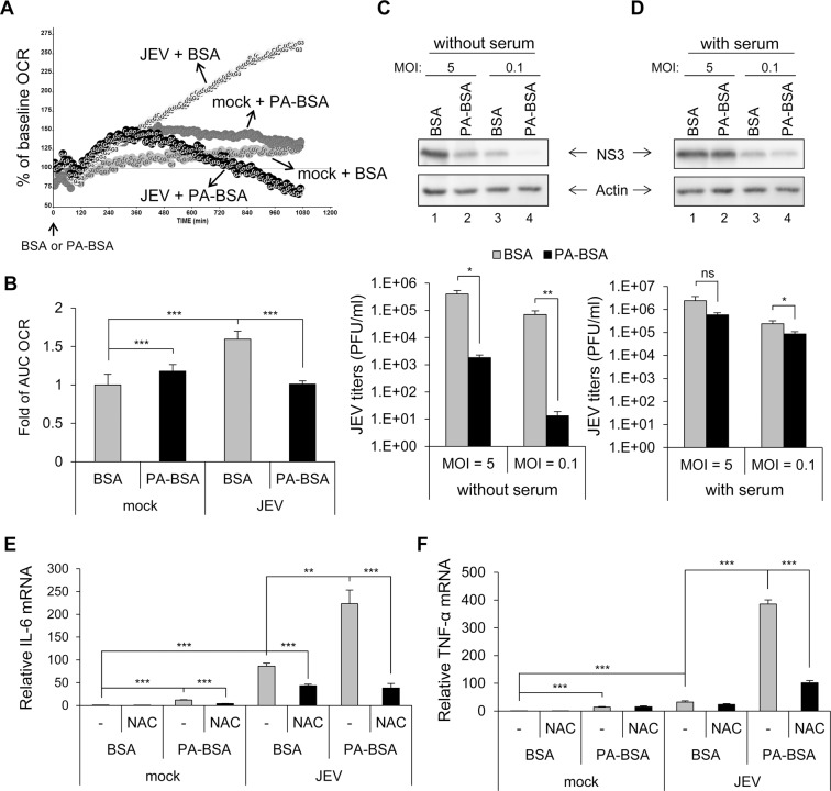
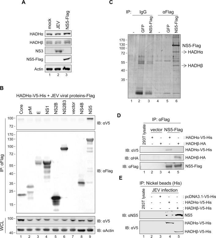
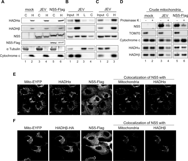
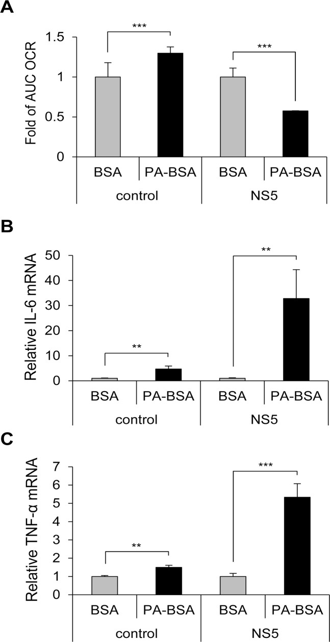
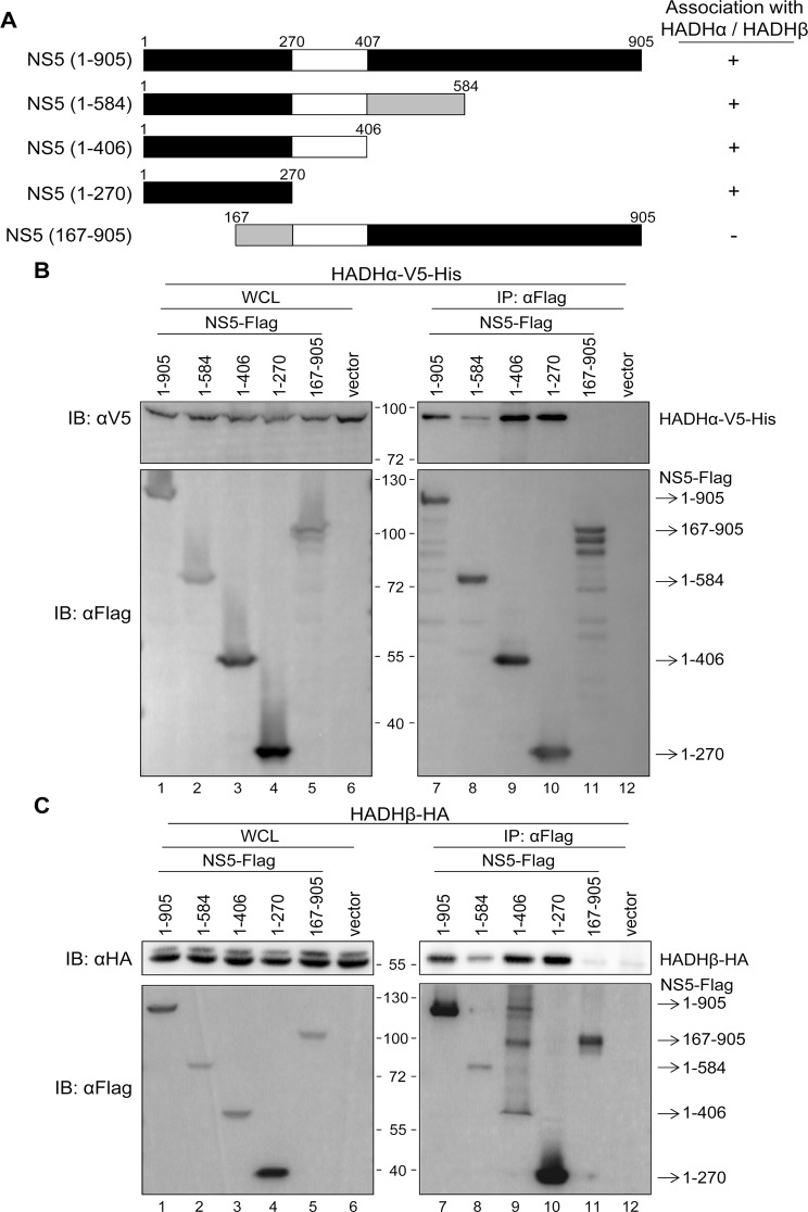
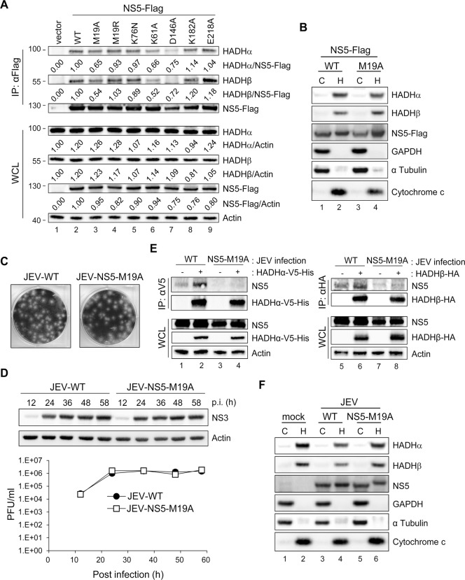
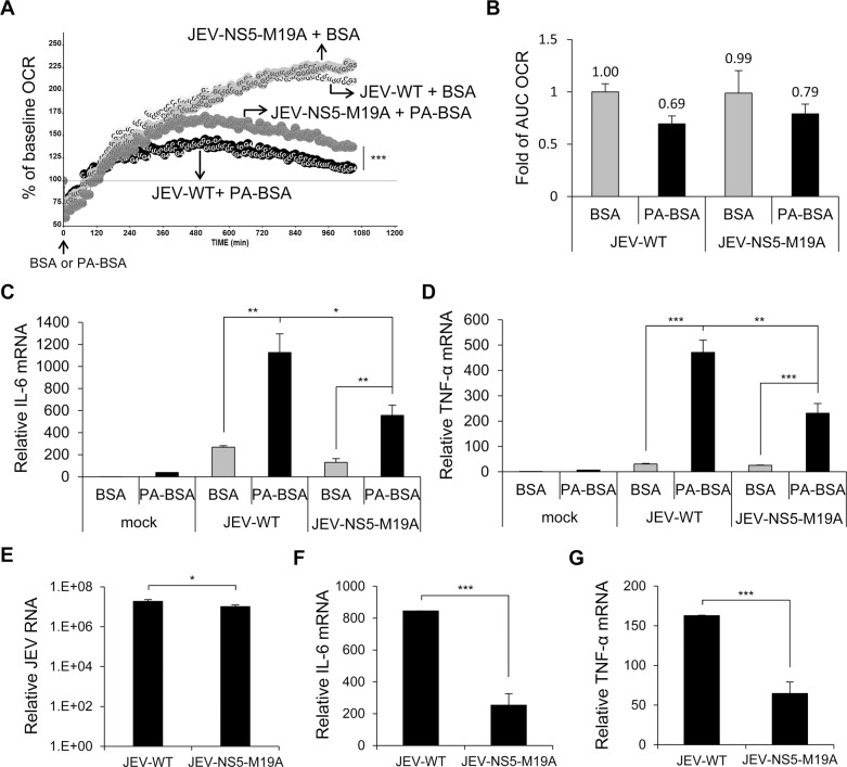
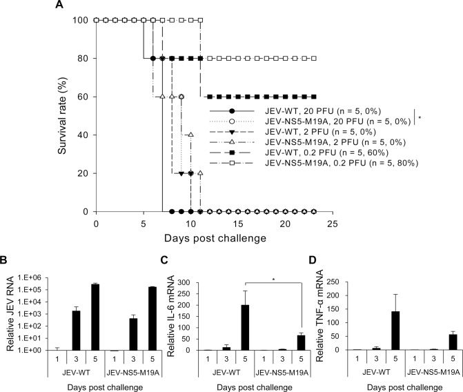
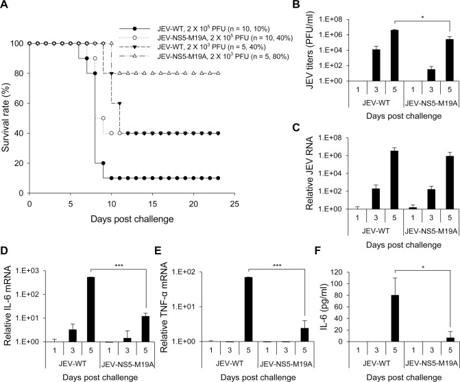
Infection with Japanese encephalitis virus (JEV) can induce the expression of pro-inflammatory cytokines and cause acute encephalitis in humans. β-oxidation breaks down fatty acids for ATP production in mitochondria, and impaired β-oxidation can induce pro-inflammatory cytokine expression. To address the role of fatty-acid β-oxidation in JEV infection, we measured the oxygen consumption rate of mock- and JEV-infected cells cultured with or without long chain fatty acid (LCFA) palmitate. Cells with JEV infection showed impaired LCFA β-oxidation and increased interleukin 6 (IL-6) and tumor necrosis factor α (TNF-α) expression. JEV nonstructural protein 5 (NS5) interacted with hydroxyacyl-CoA dehydrogenase α and β subunits, two components of the mitochondrial trifunctional protein (MTP) involved in LCFA β-oxidation, and NS5 proteins were detected in mitochondria and co-localized with MTP. LCFA β-oxidation was impaired and higher cytokines were induced in cells overexpressing NS5 protein as compared with control cells. Deletion and mutation studies showed that the N-terminus of NS5 was involved in the MTP association, and a single point mutation of NS5 residue 19 from methionine to alanine (NS5-M19A) reduced its binding ability with MTP. The recombinant JEV with NS5-M19A mutation (JEV-NS5-M19A) was less able to block LCFA β-oxidation and induced lower levels of IL-6 and TNF-α than wild-type JEV. Moreover, mice challenged with JEV-NS5-M19A showed less neurovirulence and neuroinvasiveness. We identified a novel function of JEV NS5 in viral pathogenesis by impairing LCFA β-oxidation and inducing cytokine expression by association with MTP.


Conflict of interest statement

The authors have declared that no competing interests exist.

Figures

Fig 1
Fig 1. Impaired long-chain fatty acid (LCFA) β-oxidation and induction of reactive oxygen species (ROS)-dependent pro-inflammatory cytokines in cells infected with Japanese encephalitis virus (JEV).
(A and B) A549 cells infected with JEV (multiplicity of infection [MOI] = 10) for 5 h were replenished with serum-free medium for 1 h, then treated with 200 μM palmitate conjugated to bovine serum albumin (BSA) (PA-BSA) or BSA control. (A) Real-time oxygen consumption rate (OCR) measured from 6 to 24 h post-infection (hpi). The OCR before PA-BSA or BSA treatment was set to 100%. (B) The area under the curve (AUC) OCR compared to that for mock cells treated with BSA (n = 3 per group). (C and D) A549 cells infected with JEV (MOI = 5 and 0.1) for 5 h were changed to medium without serum (C) or with serum (10% FBS) (D) for 1 h. Cells were then treated with PA-BSA or BSA for 18 h before Western blot analysis of protein levels of JEV NS3 and actin in cell lysates and virus titration in culture supernatants by plaque-forming assay (n = 3). (E and F) A549 cells were treated with N-acetylcysteine (NAC) 1 h before JEV (MOI = 10) infection and after virus adsorption. At 5 hpi, cells were incubated with serum-free medium for 1 h before treatment with PA-BSA or BSA for 18 h. RT-qPCR analysis of the relative mRNA levels of interleukin 6 (IL-6) (E) and tumor necrosis factor α (TNF-α) (F) (n = 3). Data are mean±SD. *P < 0.05, **P < 0.01, ***P < 0.001 and ns, not significant.
Fig 2
Fig 2. JEV NS5 interacts with mitochondrial trifunctional protein (MTP), the enzyme complex involved in LCFA β-oxidation.
(A) Western blot analysis of protein levels of the indicated proteins in A549 cells with JEV infection (MOI = 10) or NS5-Flag overexpression. (B) Western blot analysis of V5-tag, Flag-tag, and actin in HEK293T cells co-transfected with HADHα-V5-His plus the indicated plasmids expressing Flag-tagged JEV viral proteins for 24 h, then immunoprecipitated with anti-Flag affinity gel. WCL, whole-cell lysates. (C) Immunoprecipitation (IP) analysis with control IgG or anti-Flag affinity gel in A549, GFP-A549 and NS5-Flag-A549 cells. The protein bands identified as HADHα and HADHβ are indicated by arrows. (D) IP with anti-Flag affinity gel and Western blot analysis with the indicated antibodies in HEK293T cells co-transfected with vector control or NS5-Flag plus HADHα-V5-His or HADHβ-HA for 24 h. (E) IP analysis with nickel beads and Western blot analysis with the indicated antibodies in HEK293T cells adsorbed with JEV for 3 h, then transfected with vector control, HADHα-V5-His or HADHβ-V5-His for 24 h.
Fig 3
Fig 3. Subcellular localization of JEV NS5.
(A-C) Cellular lysates of HEK293T cells infected with JEV (MOI = 5) or transfected with NS5-Flag for 24 h underwent Qproteome Mitochondria Isolation (A) or biochemical fractionation as outlined in S5B and S5C Fig, respectively (B and C). Western blot analysis of indicated proteins in cytosolic and crude mitochondrial fractions. C, cytosolic fraction; H, heavy membrane fraction/crude mitochondrial fraction; L, light microsomal membrane fraction. (D) The crude mitochondrial fraction isolated from HEK293T cells infected with JEV (MOI = 3) or transfected with NS5-Flag for 24 h was treated with Proteinase K (100 μg/ml) for 30 min on ice. The reactants were developed by Western blot analysis with antibodies against NS5 or the indicated mitochondrial proteins. (E) Confocal microscopy of pEYFP-Mito-NS5-A549 cells stained with anti-Flag plus Alexa Fluor 568 goat anti-rabbit and anti-HADHα plus Alexa Fluor 647 goat anti-mouse antibody. (F) Confocal microscopy of pEYFP-Mito-NS5-A549 cells transfected with HADHβ-HA for 24 h and stained with anti-Flag plus Alexa Fluor 568 goat anti-rabbit and anti-HA plus Alexa Fluor 647 goat anti-mouse antibody.
Fig 4
Fig 4. Impaired LCFA β-oxidation and cytokine induction in NS5-overexpressing cells.
(A) AUC OCR for NS5-overexpressing and vector control-A549 cells incubated with serum-free medium for 1 h, then treated with PA-BSA or BSA control for 18 h (n = 2). (B and C) NS5-overexpressing and vector control-A549 cells were cultured with serum-free medium for 1 h, then incubated with PA-BSA or BSA for 24 h. RT-qPCR analysis of the relative mRNA levels of IL-6 (B) and TNF-α (C) (n = 3). Data are mean±SD. **P < 0.01, ***P < 0.001.
Fig 5
Fig 5. N-terminus of NS5 is essential for its interaction with MTP.
(A) Schematic diagram and properties of full-length and truncated NS5 constructs. (B and C) IP—Western and Western blot analysis with anti-Flag affinity gel and the indicated antibodies for Flag-tag, V5-tag and HA-tag in HEK293T cells co-transfected with full-length and truncated NS5-Flag plus HADHα-V5-His (B) or HADHβ-HA (C) for 24 h.
Fig 6
Fig 6. NS5 with mutation on residue 19 (M19A) showed reduced binding ability with MTP.
(A) Western blot analysis of indicated proteins in HEK293T cells transfected with the plasmids expressing wild type (WT)-, mutated-NS5-Flag or vector control for 24 h after immunoprecipitation with anti-Flag affinity gel. Band densities were quantified by use of MetaMorph (Molecular Devices). (B and F) HEK293T cells transfected with NS5-Flag or NS5-M19A-Flag (B) or infected with wild-type JEV (JEV-WT) or JEV-NS5-M19A (MOI = 5) (F) for 24 h underwent Qproteome Mitochondria Isolation. Western blot analysis of indicated proteins in mitochondrial and cytosolic fractions. C, cytosolic fraction; H, heavy membrane fraction/crude mitochondrial fraction. (C and D) JEV-NS5-M19A mutant virus was generated by using a JEV infectious clone. (C) Plaque morphology of wild type JEV (JEV-WT) and JEV-NS5-M19A mutant in BHK-21 cells. (D) A549 cells were infected with JEV-WT or JEV-NS5-M19A (MOI = 0.1) for the indicated times. Western blot analysis of protein levels of NS3 and actin. Plaque-forming assay of virus titration in culture supernatants (n = 3). Data are mean±SD. (E) IP analysis with V5 or HA affinity gel and Western blot analysis with the indicated antibodies in HEK293T cells adsorbed with JEV for 1 h, then transfected with HADHα-V5-His or HADHβ-HA for 24 h.
Fig 7
Fig 7. The recombinant JEV with NS5-M19A mutation is less able to block LCFA β-oxidation and induces less cytokine expression.
(A and B) A549 cells infected with JEV-WT or JEV-NS5-M19A (MOI = 10) for 5 h were changed to serum-free medium for 1 h, then incubated with PA-BSA or BSA control. (A) Real-time OCR was measured from 6 to 24 h post-infection. The OCR before PA-BSA or BSA treatment was set to 100%. (B) The AUC OCR with PA-BSA and BSA (n = 3). (C and D) A549 cells infected with the indicated JEV (MOI = 10) for 5 h were incubated with serum-free medium for 1 h before treatment with PA-BSA or BSA for 18 h. RT-qPCR analysis of relative mRNA levels of IL-6 (C) and TNF-α (D) (n = 3). (E-G) A549 cells were infected with JEV-WT or JEV-NS5-M19A (MOI = 10) for 24 h in serum (10% FBS)-containing medium. RT-qPCR analysis of relative mRNA levels of JEV RNA (E), IL-6 (F) and TNF-α (G) (n = 3). Data are mean±SD.*P < 0.05, **P < 0.01 and ***P < 0.001.
Fig 8
Fig 8. Reduced neurovirulence of NS5-M19A—mutated JEV in challenged mice.
(A) Survival in C57BL/6 mice infected with 0.2, 2 or 20 plaque-forming units (PFU) of JEV-WT or JEV-NS5-M19A by an intracerebral (i.c.) injection. The animal number (n) and survival rate for each group are shown. (B-D) RT-qPCR of relative JEV RNA (B), IL-6 (C), and TNF-α (D) mRNA levels in brain tissues of mice inoculated with JEV-WT or JEV-NS5-M19A (20 PFU) (n = 3). Data are mean±SD.*P < 0.05.
Fig 9
Fig 9. Reduced neuroinvasiveness of NS5-M19A—mutated JEV in challenged mice.
(A) Survival in C57BL/6 mice infected with 103 or 105 PFU JEV-WT or JEV-NS5-M19A by an intraperitoneal (i.p.) plus i.c. route. The animal number (n) and survival rate for each group are shown. (B) Plaque-forming assay of virus titers in brain tissues of mice inoculated with JEV-WT or JEV-NS5-M19A (105 PFU) (n = 3). (C-E) RT-qPCR of relative JEV RNA (C), IL-6 (D), and TNF-α (E) mRNA levels in brain tissues (n = 3). (F) ELISA of IL-6 protein levels in the sera samples (n = 3). Data are mean±SD.*P < 0.05, ***P < 0.001.

References

    1. Fernandez-Garcia MD, Mazzon M, Jacobs M, Amara A (2009) Pathogenesis of flavivirus infections: using and abusing the host cell. Cell Host Microbe 5: 318–328. 10.1016/j.chom.2009.04.001 - DOI - PubMed
    1. Campbell GL, Hills SL, Fischer M, Jacobson JA, Hoke CH, et al. (2011) Estimated global incidence of Japanese encephalitis: a systematic review. Bull World Health Organ 89: 766–774, 774A–774E. 10.2471/BLT.10.085233 - DOI - PMC - PubMed
    1. Le Flohic G, Porphyre V, Barbazan P, Gonzalez JP (2013) Review of climate, landscape, and viral genetics as drivers of the Japanese encephalitis virus ecology. PLoS Negl Trop Dis 7: e2208 10.1371/journal.pntd.0002208 - DOI - PMC - PubMed
    1. Mukhopadhyay S, Kuhn RJ, Rossmann MG (2005) A structural perspective of the flavivirus life cycle. Nat Rev Microbiol 3: 13–22. - PubMed
    1. Unni SK, Ruzek D, Chhatbar C, Mishra R, Johri MK, et al. (2011) Japanese encephalitis virus: from genome to infectome. Microbes Infect 13: 312–321. 10.1016/j.micinf.2011.01.002 - DOI - PubMed

Publication types

MeSH terms

Substances