. 2015 Apr 28;589(10):1080-8.
doi: 10.1016/j.febslet.2015.03.017. Epub 2015 Mar 25.

Promiscuous actions of small molecule inhibitors of the protein kinase D-class IIa HDAC axis in striated muscle

Affiliations

Promiscuous actions of small molecule inhibitors of the protein kinase D-class IIa HDAC axis in striated muscle

Douglas D Lemon et al. FEBS Lett. .

Abstract

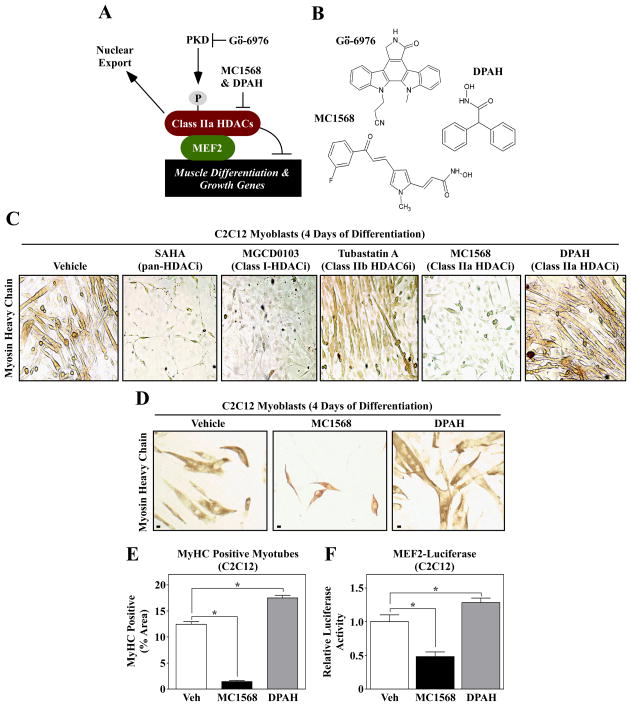
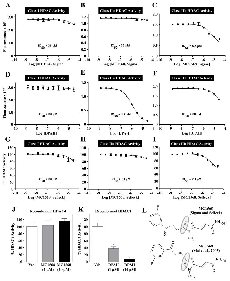
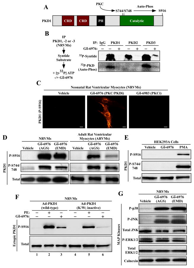
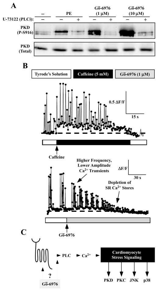
PKD-mediated phosphorylation of class IIa HDACs frees the MEF2 transcription factor to activate genes that govern muscle differentiation and growth. Studies of the regulation and function of this signaling axis have involved MC1568 and Gö-6976, which are small molecule inhibitors of class IIa HDAC and PKD catalytic activity, respectively. We describe unanticipated effects of these compounds. MC1568 failed to inhibit class IIa HDAC catalytic activity in vitro, and exerted divergent effects on skeletal muscle differentiation compared to a bona fide inhibitor of these HDACs. In cardiomyocytes, Gö-6976 triggered calcium signaling and activated stress-inducible kinases. Based on these findings, caution is warranted when employing MC1568 and Gö-6976 as pharmacological tool compounds to assess functions of class IIa HDACs and PKD.

Keywords: Class IIa histone deacetylase; Muscle; Protein kinase D; Small molecule inhibitors.


Figures

Fig. 1
Fig. 1
Differential effects of isoform-selective HDAC inhibitors on skeletal muscle differentation. (A) A schematic representation of the PKD-class IIa HDAC axis. Gö-6976 blocks the catalytic activity of PKD, while MC1568 and DPAH inhibit the enzymatic activity of class IIa HDACs. (B) The molecular structures of the compounds used in this study are shown. (C) C2C12 myoblasts were induced to differentiate in the absence (vehicle) or presence of the indicated HDAC inhibitors; SAHA (1 μM), MGCD0103 (500 nM), Tubastatin A (1 μM), MC1568 (10 μM) and DPAH (10 μM). After four days of differentiation, cells were fixed and stained with an anti-myosin antibody to reveal myosin heavy chain (MyHC)-positive skeletal myotubes. (D) C2C12 cells were induced to differentiate in the absence or presence of the class IIa HDAC inhibitors MC1568 and DPAH, stained as described for (C), and differentiation was quantified (E); Scale bar = 10 μm. (F) C2C12 myoblasts were transfected with a MEF2-dependent luciferase reporter gene, and the cells were subsequently induced to differentiate for three days in the absence or presence of MC1568 or DPAH. For (E) and (F), five independent plates of cells were analyzed per condition. Numbers represent averages +SEM; *P < 0.05 vs. vehicle-treated cells.
Fig. 2
Fig. 2
MC1568 does not block class IIa HDAC catalytic activity. Dose-response studies were performed to assess the ability of MC1568 and DPAH to inhibit HDAC catalytic activity in vitro. (A – C) MC1568 (from Sigma-Aldrich) failed to inhibit class I and class IIa HDAC activity, but did block class IIb activity at higher concentrations. (D – F) DPAH dose-dependently, and selectively, inhibited class IIa HDAC catalytic activity. MC1568 from Selleck Chemicals also failed to inhibit endogenous class IIa HDAC activity (G – I). MC1568 was unable to inhibit the catalytic activity of a recombinant form of a prototypical class IIa HDAC, HDAC4, while DPAH efficiently inhibited recombinant HDAC4 (J and K). (L) Analysis of NMR data from Sigma and Selleck revealed the commercial MC1568 to be a 2,5-disubstituted pyrrole, as opposed to the 2,4-disubstituted isomer shown in Fig. 1B.
Fig. 3
Fig. 3
Gö-6976 activates stress-dependent kinases in cardiomyocytes. (A) Schematic representation of PKD1, with cysteine-rich domains (CRD) and a pleckstrin homology (PH) domain indicated. PKC phosphorylates activation loop sites in the catalytic domain of PKD1 (serines-744 and -748), resulting in stimulation of the kinase. Serine-916 can undergo auto-phosphorylation. (B) NRVMs were stimulated with phenylephrine (PE; 10 μM) for 1 hour. Whole cell lysates were immunoprecipitated (IP) with antibodies to PKD1, -2 or -3 and incorporated into in vitro kinase reactions in the absence or presence of Gö-6976 (10 μM). Gö-6976 inhibited the ability of each PKD isoform to phosphorylate the synthetic syntide substrate, and blocked auto-phosphorylation of PKD1 and PKD3. (C) NRVMs were treated with Gö-6976 (10 μM) or Gö-6983 (10 μM) for 1 hour, and PKD serine-916 phosphorylation was assessed by indirect immunofluorescence. Scale bar = 10 μm. (D) NRVMs (left panel) or adult rat ventricular myocytes (right panel) were treated for 1 hour with vehicle control or Gö-6976 (10 μM) from two independent commercial sources, as described in the Materials and methods. Protein homogenates were immunoblotted with antibodies to the indicated forms of PKD1. (E) HEK293A fibroblasts were treated for 1 hour with Gö-6976 (10 μM) or phorbol-12-myristate-13-acetate (PMA; 50 nM), which served as a positive control for PKC and PKD activation. Immunblotting was performed as in (D). (F) NRVMs were infected with adenoviruses encoding wild-type PKD1 or PKD1 K/W, which is catalytically inactive. Cells were treated for 1 hour with PE (10 μM) or Gö-6976 (10 μM), lysed, and immunoblotted with the indicated antibodies. (G) NRVMs were treated for 1 hour with Gö-6976 (10 μM) from two different vendors, and protein homogenates were immunoblotted with antibodies to the indicated proteins.
Fig. 4
Fig. 4
Gö-6976 alters calcium signaling in cardiac myocytes. (A) NRVMs were pre-treated with the phospholipase C (PLC) inhibitor U-73122 (1 μM) for 20 minutes prior to addition of phenylephrine (PE; 10 μM) or Gö-6976 (1 or 10 μM) for 1 hour. Immunoblotting was performed to assess levels of phospho- and total PKD1. (B) NRVMs were loaded with the calcium-sensitive dye, Fluo-3 AM. Calcium fluctuations prior to and after treatment with caffeine (5 mM) or Gö-6976 (1 μM) were measured using a confocal microscope. Fluorescence amplitude data are expressed as ΔF/F, where F represents baseline fluorescence prior to application of caffeine or Gö-6976, and ΔF represents the change in peak fluorescence during the application of either compound.

References

    1. Gregoretti IV, Lee YM, Goodson HV. Molecular evolution of the histone deacetylase family: functional implications of phylogenetic analysis. J Mol Biol. 2004;338:17–31. - PubMed
    1. McKinsey TA, Zhang CL, Olson EN. Signaling chromatin to make muscle. Curr Opin Cell Biol. 2002;14:763–772. - PubMed
    1. Lemercier C, Verdel A, Galloo B, Curtet S, Brocard MP, Khochbin S. mHDA1/HDAC5 histone deacetylase interacts with and represses MEF2A transcriptional activity. J Biol Chem. 2000;275:15594–15599. - PubMed
    1. Lu J, McKinsey TA, Nicol RL, Olson EN. Signal-dependent activation of the MEF2 transcription factor by dissociation from histone deacetylases. Proc Natl Acad Sci U S A. 2000;97:4070–4075. - PMC - PubMed
    1. Miska EA, Karlsson C, Langley E, Nielsen SJ, Pines J, Kouzarides T. HDAC4 deacetylase associates with and represses the MEF2 transcription factor. EMBO J. 1999;18:5099–5107. - PMC - PubMed

Publication types

MeSH terms