. 2015 Apr 2;96(4):623-30.
doi: 10.1016/j.ajhg.2015.02.010. Epub 2015 Mar 26.

Mutations in DVL1 cause an osteosclerotic form of Robinow syndrome

Affiliations

Mutations in DVL1 cause an osteosclerotic form of Robinow syndrome

Kieran J Bunn et al. Am J Hum Genet. .

Abstract

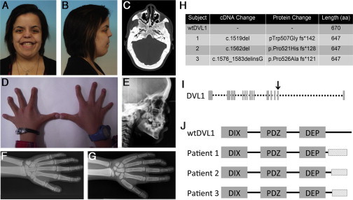
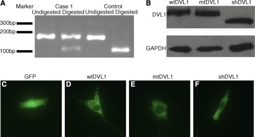
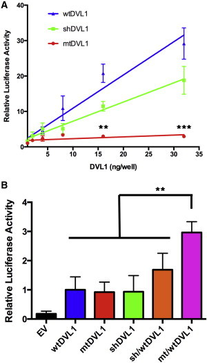
Robinow syndrome (RS) is a phenotypically and genetically heterogeneous condition that can be caused by mutations in genes encoding components of the non-canonical Wnt signaling pathway. In contrast, germline mutations that act to increase canonical Wnt signaling lead to distinctive osteosclerotic phenotypes. Here, we identified de novo frameshift mutations in DVL1, a mediator of both canonical and non-canonical Wnt signaling, as the cause of RS-OS, an RS subtype involving osteosclerosis, in three unrelated individuals. The mutations all delete the DVL1 C terminus and replace it, in each instance, with a novel, highly basic sequence. We showed the presence of mutant transcript in fibroblasts from one individual with RS-OS and demonstrated unimpaired protein stability with transfected GFP-tagged constructs bearing a frameshift mutation. In vitro TOPFlash assays, in apparent contradiction to the osteosclerotic phenotype, revealed that the mutant allele was less active than the wild-type allele in the canonical Wnt signaling pathway. However, when the mutant and wild-type alleles were co-expressed, canonical Wnt activity was 2-fold higher than that in the wild-type construct alone. This work establishes that DVL1 mutations cause a specific RS subtype, RS-OS, and that the osteosclerosis associated with this subtype might be the result of an interaction between the wild-type and mutant alleles and thus lead to elevated canonical Wnt signaling.


Figures

Figure 1
Figure 1
Clinical Presentation of and Mutations in RS-OS (A–G) Clinical features of RS-OS. (A and B) Facial appearance of subject 1. Note the midface hypoplasia, flat facial profile, hypertelorism, and broad mouth. (C) Transverse CT of subject 1. Note the pronounced osteosclerosis of the cranial vault. (D) Appearance of the hands of subject 2. Note the the camptodactyly and brachydactyly. (E) Lateral X-ray of the skull of subject 2. Note the thickened calvarium and increased bone density. (F) X-ray of the left hand, wrist, and forearm of subject 1. Note the osteosclerosis in the cortices of the long bones of the forearm and the camptodactyly, clinodactyly, and bifid thumb. (This image was reproduced with permission from Bunn et al.17) (G) X-ray of the left hand, wrist, and forearm of subject 1. Note the cortical osteosclerosis of the forearm and the camptodactyly, clinodactyly, and bifid thumb (less obvious than in subject 1). (H) DVL1 mutations leading to RS-OS. (I) DVL1 structure showing exons. The arrow indicates exon 14, the location of the three mutations. (J) An illustration of DVL1 shows the similarity among the altered proteins and their difference from the wild-type. The shaded box indicates the novel shared C-terminal sequence.
Figure 2
Figure 2
Expression and Localization of Frameshifted and Truncated DVL1 (A) The DVL1 PCR product was digested with the restriction enzyme BstN1 for 4 hr at 60°C. The digest shows the presence of mutant transcript (which is refractory to digestion) alongside wild-type transcript in fibroblasts obtained from subject 1. (B) Chemiluminescent immunoblot of C2C12 cells (40,000/well) transiently transfected (0.6 μl/well of Lipofectamine2000, Life Technologies) with EGFP-tagged DVL1 constructs (100 ng/well,incubated for 24 hr) demonstrates comparable protein levels (anti-GFP, A6455, Life Technologies; anti-GAPDH, G8795, Sigma). Note the size difference between the truncated DVL and the other constructs. (C–F) Representative images from fluorescent microscopy of C2C12 cells transiently transfected with EGFP or the EGFP-DVL1 constructs (100 ng/well) show that p.Trp507 DVL1 (shDVL1) and mtDVL1 retain the ability to form puncta. shDVL1 is a construct that leads to a truncated protein terminating at the site of the frameshift mutation (c.1519insTAA [p.Trp507]) found in subject 1.
Figure 3
Figure 3
Impact of DVL1 Constructs on Canonical Wnt Signaling (A) TOPFlash reporter assay in C2C12 cells. Cells were transiently transfected with 80 ng/well of TOPFlash reporter, 20 ng/well of a constitutively active β-galactosidase construct, and a variable amount of a DVL1 construct and incubated for 18 hr. Combined luciferase activities of three independent experiments normalized to the β-galactosidase activity are depicted and reported as a relative increase over that of an empty vector (n = 3). Error bars represent the SEM. Log-transformed two-way ANOVA found a significant difference between the mutant and every other construct (p < 0.001). Individual p values were calculated with Tukey HSD tests and denote the difference between mtDVL1 and every other construct. (B) C2C12 cells were transiently transfected with a fixed amount of each DVL1 construct (4 ng/well) or with a 1:1 stoichiometric ratio of two constructs with the same total amount of DVL1 and incubated for 18 hr. The same reporters and processing were used as above, and the luciferase activity is expressed as a proportion of the wtDVL1 activation (n = 5). Error bars represent the SEM. P values were calculated with Tukey HSD tests (p 0.05, ∗∗p = 0.01, ∗∗∗p = 0.001).

References

    1. Mazzeu J.F., Pardono E., Vianna-Morgante A.M., Richieri-Costa A., Ae Kim C., Brunoni D., Martelli L., de Andrade C.E., Colin G., Otto P.A. Clinical characterization of autosomal dominant and recessive variants of Robinow syndrome. Am. J. Med. Genet. A. 2007;143:320–325. - PubMed
    1. Afzal A.R., Rajab A., Fenske C.D., Oldridge M., Elanko N., Ternes-Pereira E., Tüysüz B., Murday V.A., Patton M.A., Wilkie A.O., Jeffery S. Recessive Robinow syndrome, allelic to dominant brachydactyly type B, is caused by mutation of ROR2. Nat. Genet. 2000;25:419–422. - PubMed
    1. van Bokhoven H., Celli J., Kayserili H., van Beusekom E., Balci S., Brussel W., Skovby F., Kerr B., Percin E.F., Akarsu N., Brunner H.G. Mutation of the gene encoding the ROR2 tyrosine kinase causes autosomal recessive Robinow syndrome. Nat. Genet. 2000;25:423–426. - PubMed
    1. Person A.D., Beiraghi S., Sieben C.M., Hermanson S., Neumann A.N., Robu M.E., Schleiffarth J.R., Billington C.J., Jr., van Bokhoven H., Hoogeboom J.M. WNT5A mutations in patients with autosomal dominant Robinow syndrome. Dev. Dyn. 2010;239:327–337. - PMC - PubMed
    1. Niehrs C. The complex world of WNT receptor signalling. Nat. Rev. Mol. Cell Biol. 2012;13:767–779. - PubMed

Publication types

MeSH terms

Supplementary concepts

LinkOut - more resources