. 2015 Apr 2;96(4):565-80.
doi: 10.1016/j.ajhg.2015.02.009. Epub 2015 Mar 26.

A large-scale genetic analysis reveals a strong contribution of the HLA class II region to giant cell arteritis susceptibility

F David Carmona  1 Sarah L Mackie  2 Jose-Ezequiel Martín  3 John C Taylor  4 Augusto Vaglio  5 Stephen Eyre  6 Lara Bossini-Castillo  3 Santos Castañeda  7 Maria C Cid  8 José Hernández-Rodríguez  8 Sergio Prieto-González  8 Roser Solans  9 Marc Ramentol-Sintas  9 M Francisca González-Escribano  10 Lourdes Ortiz-Fernández  10 Inmaculada C Morado  11 Javier Narváez  12 José A Miranda-Filloy  13 Spanish GCA GroupLorenzo Beretta  14 Claudio Lunardi  15 Marco A Cimmino  16 Davide Gianfreda  17 Daniele Santilli  18 Giuseppe A Ramirez  19 Alessandra Soriano  20 Francesco Muratore  21 Giulia Pazzola  21 Olga Addimanda  21 Cisca Wijmenga  22 Torsten Witte  23 Jan H Schirmer  24 Frank Moosig  24 Verena Schönau  25 Andre Franke  26 Øyvind Palm  27 Øyvind Molberg  27 Andreas P Diamantopoulos  28 Simon Carette  29 David Cuthbertson  30 Lindsy J Forbess  31 Gary S Hoffman  32 Nader A Khalidi  33 Curry L Koening  34 Carol A Langford  32 Carol A McAlear  35 Larry Moreland  36 Paul A Monach  37 Christian Pagnoux  29 Philip Seo  38 Robert Spiera  39 Antoine G Sreih  35 Kenneth J Warrington  40 Steven R Ytterberg  40 Peter K Gregersen  41 Colin T Pease  42 Andrew Gough  43 Michael Green  44 Lesley Hordon  45 Stephen Jarrett  46 Richard Watts  47 Sarah Levy  48 Yusuf Patel  49 Sanjeet Kamath  50 Bhaskar Dasgupta  51 Jane Worthington  6 Bobby P C Koeleman  52 Paul I W de Bakker  53 Jennifer H Barrett  4 Carlo Salvarani  21 Peter A Merkel  35 Miguel A González-Gay  54 Ann W Morgan  4 Javier Martín  3
Collaborators, Affiliations

A large-scale genetic analysis reveals a strong contribution of the HLA class II region to giant cell arteritis susceptibility

F David Carmona et al. Am J Hum Genet. .

Abstract

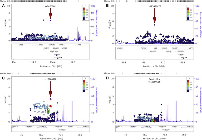
We conducted a large-scale genetic analysis on giant cell arteritis (GCA), a polygenic immune-mediated vasculitis. A case-control cohort, comprising 1,651 case subjects with GCA and 15,306 unrelated control subjects from six different countries of European ancestry, was genotyped by the Immunochip array. We also imputed HLA data with a previously validated imputation method to perform a more comprehensive analysis of this genomic region. The strongest association signals were observed in the HLA region, with rs477515 representing the highest peak (p = 4.05 × 10(-40), OR = 1.73). A multivariate model including class II amino acids of HLA-DRβ1 and HLA-DQα1 and one class I amino acid of HLA-B explained most of the HLA association with GCA, consistent with previously reported associations of classical HLA alleles like HLA-DRB1(∗)04. An omnibus test on polymorphic amino acid positions highlighted DRβ1 13 (p = 4.08 × 10(-43)) and HLA-DQα1 47 (p = 4.02 × 10(-46)), 56, and 76 (both p = 1.84 × 10(-45)) as relevant positions for disease susceptibility. Outside the HLA region, the most significant loci included PTPN22 (rs2476601, p = 1.73 × 10(-6), OR = 1.38), LRRC32 (rs10160518, p = 4.39 × 10(-6), OR = 1.20), and REL (rs115674477, p = 1.10 × 10(-5), OR = 1.63). Our study provides evidence of a strong contribution of HLA class I and II molecules to susceptibility to GCA. In the non-HLA region, we confirmed a key role for the functional PTPN22 rs2476601 variant and proposed other putative risk loci for GCA involved in Th1, Th17, and Treg cell function.


Figures

Figure 1
Figure 1
Manhattan Plot Representation of the Immunochip Results The −log10 of the combined logistic regression test p values are plotted against their physical chromosomal position. The red line represents the genome-wide level of significance (p < 5 × 10−8), and the blue line represents the statistical significance for suggestive signals (p < 1 × 10−4). The most relevant associations are highlighted.
Figure 2
Figure 2
Manhattan Plot Representation of the Step-wise Conditional Logistic Regression of the HLA Region (A) Unconditioned test of the HLA region. (B) Results of the HLA region after conditioning to HLA-DRβ1 His13. (C) Results of the HLA region after conditioning to HLA-DRβ1 His13 and HLA-DQα1 Gly56. (D) Results of the HLA region after conditioning to HLA-DRβ1 His13, HLA-DQα1 Gly56, and HLA-B Thr45. The −log10 of the combined logistic regression test p values are plotted against their physical chromosomal position. A red/blue color gradient was used to represent the effect size of each analyzed variant (red for risk and blue for protection). The red line represents the genome-wide level of significance (p < 5 × 10−8).
Figure 3
Figure 3
Haplotypes Observed in Our Dataset between the Residues of the Class II Positions of the Model that Best Explains the Association of This Genomic Region with Giant Cell Arteritis (A) Graphical representation of the four haplotypes formed by the amino acids of the DQα1 positions 47, 56, and 76. The frequency of every amino acid is shown in brackets. (B) Each row refers to the haplotype of one HLA-DRβ1 13 amino acid and one HLA-DQα1 haplotype. Haplotype frequencies of case subjects (red) and control subjects (blue), odds ratios (OR), and −log10 of the logistic regression test p values are plotted. A red/blue color gradient was used in the OR bars to represent the effect size of each analyzed variant (red for risk and blue for protection).
Figure 4
Figure 4
Regional Plots of the Three Most Associated Loci with GCA outside the HLA Region in the Overall Meta-analysis after Imputation (A) Protein tyrosine phosphatase non-receptor type 22 (PTPN22) region. (B) v-rel avian reticuloendotheliosis viral oncogene homolog (REL) region. (C) Leucine-rich repeat containing 32 (LRRC32) region. (D) Results for the LRRC32 region after conditioning for the lead SNP (rs10160518). Red arrows point to the lead variants (highlighted in violet).
Figure 5
Figure 5
Distributions of Genetic Risk Score Predictive for Susceptibility to Develop Rheumatoid Arthritis by Disease Status (A, C, and E) Histogram of genetic risk scores calculated with all the RA alleles (A), with non-HLA alleles only (C), or with HLA alleles only (E). (B, D, and F) Distribution curve of genetic risk scores calculated with all the RA alleles (B), with non-HLA alleles only (D), or with HLA alleles only (F).
Figure 6
Figure 6
Ribbon Representation of the HLA Molecules HLA-DR, HLA-DQ, and HLA-B/β2 Microglobulin The amino acid positions of the HLA model associated with GCA are highlighted in orange.

References

    1. Jennette J.C., Falk R.J., Bacon P.A., Basu N., Cid M.C., Ferrario F., Flores-Suarez L.F., Gross W.L., Guillevin L., Hagen E.C. 2012 revised International Chapel Hill Consensus Conference Nomenclature of Vasculitides. Arthritis Rheum. 2013;65:1–11. - PubMed
    1. Gonzalez-Gay M.A., Vazquez-Rodriguez T.R., Lopez-Diaz M.J., Miranda-Filloy J.A., Gonzalez-Juanatey C., Martin J., Llorca J. Epidemiology of giant cell arteritis and polymyalgia rheumatica. Arthritis Rheum. 2009;61:1454–1461. - PubMed
    1. González-Gay M.A., Blanco R., Rodríguez-Valverde V., Martínez-Taboada V.M., Delgado-Rodriguez M., Figueroa M., Uriarte E. Permanent visual loss and cerebrovascular accidents in giant cell arteritis: predictors and response to treatment. Arthritis Rheum. 1998;41:1497–1504. - PubMed
    1. Ly K.H., Régent A., Tamby M.C., Mouthon L. Pathogenesis of giant cell arteritis: More than just an inflammatory condition? Autoimmun. Rev. 2010;9:635–645. - PubMed
    1. Carmona F.D., González-Gay M.A., Martín J. Genetic component of giant cell arteritis. Rheumatology (Oxford) 2014;53:6–18. - PubMed

Publication types