

Pathogenicity and pathogenesis of a United States porcine deltacoronavirus cell culture isolate in 5-day-old neonatal piglets
- PMID: 25817405
- PMCID: PMC7111688
- DOI: 10.1016/j.virol.2015.03.024
Pathogenicity and pathogenesis of a United States porcine deltacoronavirus cell culture isolate in 5-day-old neonatal piglets
Abstract
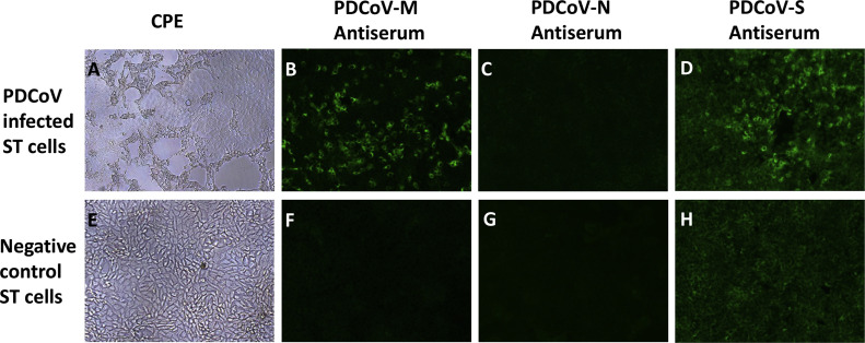
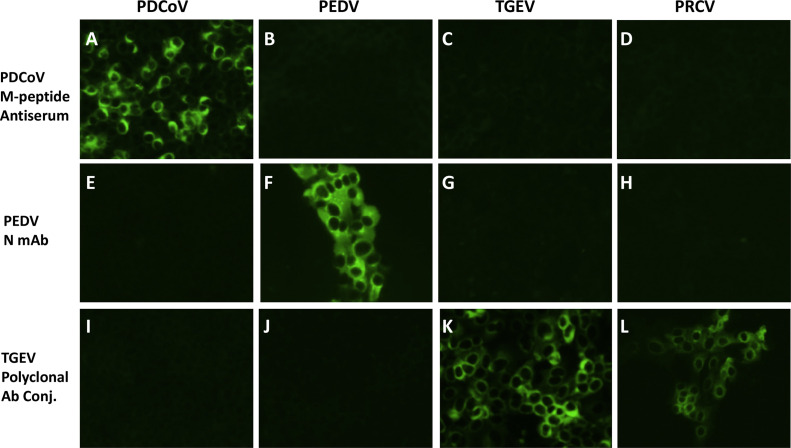
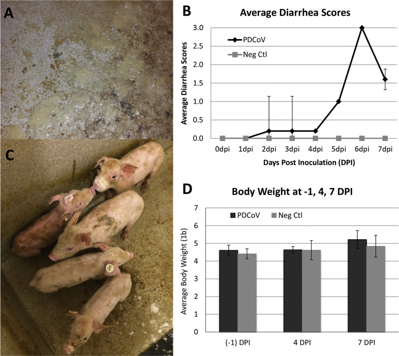
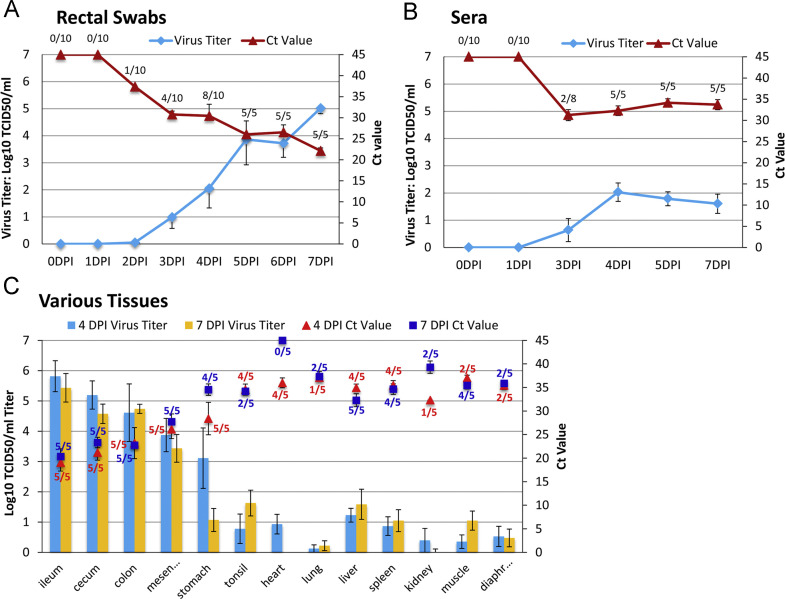
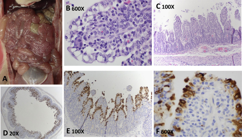
Porcine deltacoronavirus (PDCoV) was first identified in Hong Kong in 2009-2010 and reported in United States swine for the first time in February 2014. However, diagnostic tools other than polymerase chain reaction for PDCoV detection were lacking and Koch's postulates had not been fulfilled to confirm the pathogenic potential of PDCoV. In the present study, PDCoV peptide-specific rabbit antisera were developed and used in immunofluorescence and immunohistochemistry assays to assist PDCoV diagnostics. The pathogenicity and pathogenesis of PDCoV was investigated following orogastric inoculation of 5-day-old piglets with a plaque-purified PDCoV cell culture isolate (3 × 10(4) TCID50 per pig). The PDCoV-inoculated piglets developed mild to moderate diarrhea, shed increasing amount of virus in rectal swabs from 2 to 7 days post inoculation, and developed macroscopic and microscopic lesions in small intestines with viral antigen confirmed by immunohistochemistry staining. This study experimentally confirmed PDCoV pathogenicity and characterized PDCoV pathogenesis in neonatal piglets.
Keywords: Atrophic enteritis; Coronavirus; Immunohistochemistry; PDCoV; Peptide-specific antisera.
Copyright © 2015 Elsevier Inc. All rights reserved.
Figures
References
-
- Chen Q., Li G., Stasko J., Thomas J.T., Stensland W.R., Pillatzki A.E., Gauger P.C., Schwartz K.J., Madson D., Yoon K.J., Stevenson G.W., Burrough E.R., Harmon K.M., Main R.G., Zhang J. Isolation and characterization of porcine epidemic diarrhea viruses associated with the 2013 disease outbreak among swine in the United States. J. Clin. Microbiol. 2014;52(1):234–243. - PMC - PubMed
-
- Debouck P., Pensaert M., Coussement W. The pathogenesis of an enteric infection in pigs, experimentally induced by the coronavirus-like agent, CV777. Vet. Microbiol. 1981;6:157–165.
-
- Dong B.Q., Liu W., Fan X.H., Vijaykrishna D., Tang X.C., Gao F., Li L.F., Li G.J., Zhang J.X., Yang L.Q., Poon L.L., Zhang S.Y., Peiris J.S., Smith G.J., Chen H., Guan Y. Detection of a novel and highly divergent coronavirus from asian leopard cats and Chinese ferret badgers in Southern China. J. Virol. 2007;81(13):6920–6926. - PMC - PubMed
-
- Kim B., Chae C. Experimental infection of piglets with transmissible gastroenteritis virus: a comparison of three strains (Korean, Purdue and Miller) J. Comp. Pathol. 2002;126(1):30–37. - PubMed
Publication types
MeSH terms
Substances
Associated data
- Actions
LinkOut - more resources
Full Text Sources
Other Literature Sources
