

Morphologic changes in the mesolimbic pathway in Parkinson's disease motor subtypes
- PMID: 25817514
- PMCID: PMC4424152
- DOI: 10.1016/j.parkreldis.2015.03.008
Morphologic changes in the mesolimbic pathway in Parkinson's disease motor subtypes
Abstract
Background: Parkinson's disease (PD) is a common neurodegenerative disorder associated with gray matter atrophy. Cortical atrophy patterns may further help distinguish between PD motor subtypes. Comparable differences in subcortical volumes have not been found.
Methods: Twenty-one cognitively intact and treated PD patients, including 12 tremor dominant (TD) subtype, Nine postural instability gait dominant (PIGD) subtype, and 20 matched healthy control subjects underwent 3.0 T high-resolution structural MRI scanning. Subcortical volumetric analysis was performed using FreeSurfer and shape analysis was performed with FIRST to assess for differences between PD patients and controls and between PD subtypes.
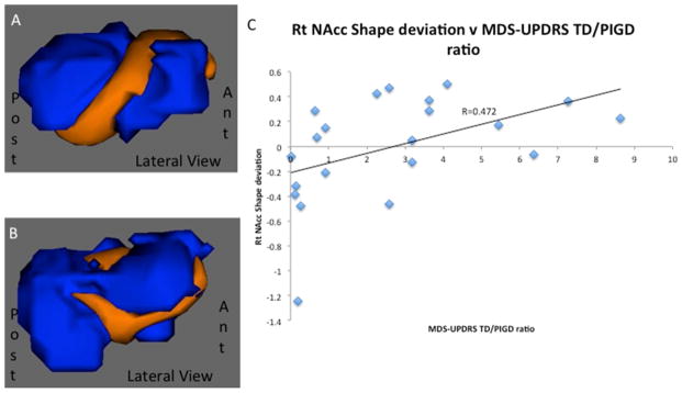
Results: No significant differences in subcortical volumes were found between motor PD subtypes, but comparing grouped PD patients with controls revealed a significant increase in hippocampal volume in PD patients (p = 0.03). A significant shape difference was detected in the right nucleus accumbens (NAcc) between PD and controls and between motor subtypes. Shape differences were driven by positive deviations in the TD subtype. Correlation analysis revealed a trend between hippocampal volume and decreasing MDS-UPDRS (p = 0.06).
Conclusion: While no significant differences in subcortical volumes between PD motor subtypes were found, increased hippocampal volumes were observed in PD patients compared to controls. Right NAcc shape differences in PD patients were driven by changes in the TD subtype. These unexpected findings may be related to the effects of chronic dopaminergic replacement on the mesolimbic pathway. Further studies are needed to replicate and determine the clinical significance of such morphologic changes.
Keywords: Dopamine; Hippocampus; Nucleus accumbens; Parkinson's disease; Shape analysis; Volumetric analysis.
Copyright © 2015 Elsevier Ltd. All rights reserved.
Conflict of interest statement
Financial disclosures and Conflicts of interest: None
Figures
References
-
- Selikhova M, et al. A clinico-pathological study of subtypes in Parkinson’s disease. Brain. 2009;132:2947–2957. - PubMed
-
- Eggers C, Kahraman D, Fink GR, Schmidt M, Timmermann L. Akinetic-rigid and tremor-dominant Parkinson’s disease patients show different patterns of FP-CIT Single photon emission computed tomography. Mov Disord. 2011;26:416–423. - PubMed
-
- Linder J, et al. Degenerative changes were common in brain magnetic resonance imaging in patients with newly diagnosed Parkinson’s disease in a population-based cohort. J Neurol. 2009;256:1671–1680. - PubMed
Publication types
MeSH terms
Grants and funding
LinkOut - more resources
Full Text Sources
Other Literature Sources
Medical
Research Materials
Miscellaneous
