. 2015 Aug;63(2):388-98.
doi: 10.1016/j.jhep.2015.03.021. Epub 2015 Mar 25.

Pivotal preclinical trial of the spheroid reservoir bioartificial liver

Affiliations

Pivotal preclinical trial of the spheroid reservoir bioartificial liver

Jaime M Glorioso et al. J Hepatol. 2015 Aug.

Abstract

Background & aims: The neuroprotective effect of the spheroid reservoir bioartificial liver (SRBAL) was evaluated in a porcine model of drug-overdose acute liver failure (ALF).

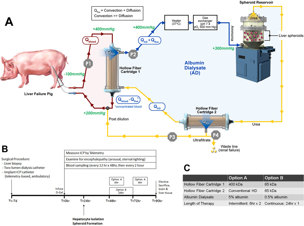
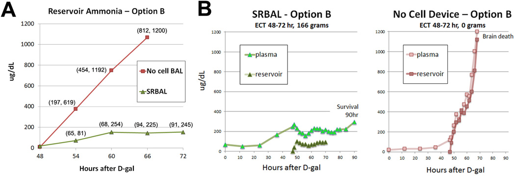
Methods: Healthy pigs were randomized into three groups (standard therapy (ST) alone, ST+No-cell device, ST+SRBAL device) before placement of an implantable intracranial pressure (ICP) monitor and a tunneled central venous catheter. One week later, pigs received bolus infusion of the hepatotoxin D-galactosamine and were followed for up to 90h.

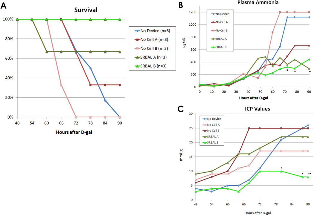
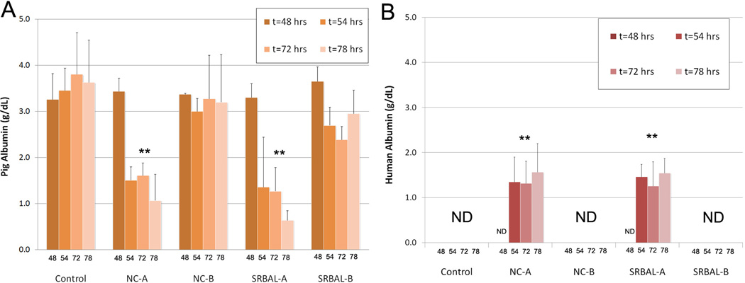
Results: At 48h, all animals had developed encephalopathy and biochemical changes confirming ALF; extracorporeal treatment was initiated and pigs were observed up to 90h after drug infusion. Pigs treated with the SRBAL, loaded with porcine hepatocyte spheroids, had improved survival (83%, n=6) compared to ST alone (0%, n=6, p=0.003) and No-cell device therapy (17%, n=6, p=0.02). Ammonia detoxification, peak levels of serum ammonia and peak ICP, and pig survival were influenced by hepatocyte cell dose, membrane pore size and duration of SRBAL treatment. Hepatocyte spheroids remained highly functional with no decline in mean oxygen consumption from initiation to completion of treatment.

Conclusions: The SRBAL improved survival in an allogeneic model of drug-overdose ALF. Survival correlated with ammonia detoxification and ICP lowering indicating that hepatocyte spheroids prevented the cerebral manifestations of ALF (brain swelling, herniation, death). Further investigation of SRBAL therapy in a clinical setting is warranted.

Keywords: Bioartificial liver; D-galactosamine; Hepatocyte; Liver failure; Spheroid.


Figures

Fig. 1
Fig. 1. Experimental Design
Panel A – Schematic of Extracorporeal Circuit including SRBAL Apparatus. Red line indicates the blood compartment, while yellow/blue line indicates the acellular albumin dialysate (AD) compartment. Flow rates in these two compartments, Qblood and QAD, along with flux across the membrane of Hollow Fiber Cartridge 1, Qflux, and waste line flow were determined by pumps P1, P2, P3, and P4. Standard flow rates for the system were Qblood = 200 mL/min, QAD = 200 mL/min, Qflux. = 50 mL/min. The waste line pump (P4) was not operated in this study (waste line flow = 0mL/min). Pressure limits are labeled in green italic font. Removal of permeable waste molecules, such as ammonia, from the blood occurred by both diffusion and convection (35). Blue lines indicate areas of high ammonia concentration in the AD circuit where total flow rate was QAD+ Qflux. The Spheroid Reservoir functioned as a suspension bioreactor with fluid entering its bottom and exiting its top. Oxygen consumption by hepatocyte spheroids was determined by the product of QAD+ Qflux multiplied by the difference in oxygen tension between inlet and outlet. An oxygenator was used to maintain the inlet oxygen tension above 500 mmHg. Exit oxygen tension averaged about 100mmHg. An outflow filter (0.2µm) prevented loss of hepatocytes from the reservoir. The semipermeable membrane of Hollow Fiber Cartridge 2 was used to maximize removal of detoxification products, such as urea, and achieve fluid balance in the AD circuit. Panel B – Experimental time line showing the sequence of events and timing of extracorporeal treatments of Option A and Option B. Panel C – detailed summary of Option A and Option B.
Fig. 2
Fig. 2. SRBAL Therapy improved Survival, Plasma Ammonia, Intracranial Pressure (ICP)
Panel A – Survival curves of all five treatment conditions. Survival at 90 hours of pigs treated with the SRBAL (Option A and Option B) was significantly improved (83%, n=6) compared to the two control groups, No-cell device therapy (17%, n=6, p=0.01) and ST alone (0%, n=6, p=0.004) groups. 100% survival was observed under conditions of SRBAL Option B. Panel B - Median values of plasma ammonia for each of the five treatment conditions are shown. Pigs treated with the SRBAL (both Option A and Option B) had stabilization in plasma ammonia during therapy, beginning at 48 hr, (* indicates p<0.05 compared to the ST control group at the corresponding time point). Panel C - Median ICP values for pigs in each of the five treatment conditions are shown. Pigs treated with the SRBAL Option B had lower final ICP compared to the ST group (** p<0.005). The difference in ICP of pigs in SRBAL Option B and ST control groups was significant from 80–90 hours (* p<0.05).
Fig. 3
Fig. 3. Porcine and Human Albumin in Pig Plasma
Panels A – Levels of porcine albumin declined significantly in pig blood during ECT therapy under Option A conditions. Bars equal mean values, error bars equal standard deviation. ** indicates p<0.05 vs. corresponding 48hr time point. Panels B – Levels of human albumin rose significantly in pig blood during ECT therapy under Option A conditions. Bars equal mean values, error bars equal standard deviation. ** indicates p<0.05 vs. corresponding 48hr time point. ND indicates level less than 0.1g/dL.
Fig. 4
Fig. 4. Liver Histology
Panels A/E – normal liver at baseline stained with H&E/Ki67. Panels B/C/D – ALF liver histology stained with H&E at increasing magnification demonstrates extensive hemorrhagic necrosis (red arrows), bile duct proliferation (green arrows), and inflammation (yellow arrows). Panel F – Ki67 staining was increased from baseline indicating a strong proliferative response (green arrows) of both hepatocytes and bile duct cells (see inset) to acute liver injury. Scale bars indicate relative size as marked.
Fig. 5
Fig. 5. Ammonia Metabolism by Hepatocyte Spheroids
Panel A – Ammonia levels in the reservoir compartment during ECT – Option B conditions. Rate of ammonia detoxification was increased significantly (p=0.003) by spheroid hepatocytes in SRBAL compared to No-cell conditions (Option B conditions, n=3 treatments per group). Markers indicate median values (min, max) at each time point. Panel B –Plasma and reservoir levels of ammonia, obtained simultaneously, from representative animals treated under cellular (SRBAL Option B, left) and no cell (ST+No cell Device, right) device configurations. Ammonia concentration remained stable in the reservoir of the device loaded with 166 grams of hepatocyte spheroids and the plasma of its treatment animal. In contrast, under the same time frame and similar starting conditions at t=48hrs, ammonia concentrations rose sharply in the no-cell reservoir and plasma of its treatment animal until reaching a study endpoint (brain death). Both devices started treatment with minimal detectable ammonia in the reservoir circuit (t=48hr).

Comment in

References

    1. Lee WM, Squires RH, Jr, Nyberg SL, Doo E, Hoofnagle JH. Acute liver failure: Summary of a workshop. Hepatology. 2008;47:1401–1415. - PMC - PubMed
    1. Kjaergard L, Liu J, Als-Nielsen B, al e. Artificial and bioartificial support systems for acute and acute-on-chronic liver failure: a systematic review. JAMA. 2003;289:217–222. - PubMed
    1. Demetriou AA, Brown RS, Jr, Busuttil RW, Fair J, McGuire BM, Rosenthal P, Am Esch JS, 2nd, et al. Prospective, randomized, multicenter, controlled trial of a bioartificial liver in treating acute liver failure. Ann Surg. 2004;239:660–667. discussion 667–670. - PMC - PubMed
    1. Saliba F, Camus C, Durand F, Mathurin P, Letierce A, Delafosse B, Barange K, et al. Albumin dialysis with a noncell artificial liver support device in patients with acute liver failure: a randomized controlled trial. Ann Intern Med. 2013;159:522–531. - PubMed
    1. Yu Y, Fisher JE, Lillegard JB, Rodysill B, Amiot B, Nyberg SL. Cell therapies for liver diseases. Liver transplantation. 2012;18:9–21. - PMC - PubMed

Publication types