. 2015 Mar-Apr;29(2):499-504.
doi: 10.1111/jvim.12538.

Hypercoagulability in dogs with blastomycosis

Affiliations

Hypercoagulability in dogs with blastomycosis

M A McMichael et al. J Vet Intern Med. 2015 Mar-Apr.

Abstract

Background: Blastomycosis is a potentially fatal fungal disease that most commonly affects humans and dogs. The organism causes systemic inflammation and has a predilection for the lungs. The inflammation might lead to a hypercoagulable state with microemboli in the pulmonary circulation which could contribute to inadequate oxygen exchange in infected dogs.

Hypothesis/objectives: Dogs with blastomycosis will be hypercoagulable compared with healthy case-matched controls.

Animals: Client-owned dogs with a diagnosis of blastomycosis (n = 23) and healthy case-matched controls (n = 23).

Methods: Prospective case-controlled study of client-owned dogs presented to a veterinary teaching hospital with clinical signs compatible with blastomycosis. Complete blood counts, fibrinogen, PT, aPTT, thromboelastometry (TE), thrombin antithrombin complexes (TAT), and thrombin generation were evaluated.

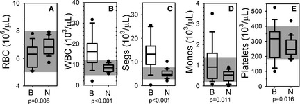
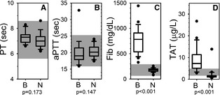
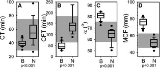
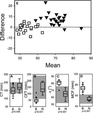
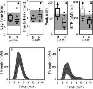
Results: Cases had a leukocytosis compared with controls [mean (SD) 16.6 (7.6) × 10(3)/μL versus 8.2 (1.8) × 10(3)/μL, P < .001], hyperfibrinogenemia [median 784 mg/dL, range 329-1,443 versus median 178 mg/dL, range 82-257, P < .001], and increased TAT concentrations [mean (SD) 9.0 (5.7) μg/L versus 2.0 (2.8) μg/L, P < .001]. As compared to controls, cases were also hypercoagulable as evaluated by thromboelastometry and had increased in vitro thrombin generation on calibrated automated thrombography.

Conclusions and clinical importance: Hypercoagulability occurs in dogs with systemic blastomycosis. Additional studies are needed to explore a possible contribution of thrombogenicity to the clinical manifestations of systemic blastomycosis.

Keywords: Canine; Thromboelastography; Thromboelastometry; Thrombosis.


Figures

Figure 1
Figure 1
Blood cells. Box and whisker plots describing results of complete blood counts for the blastomycosis cohort (“B”) and the normal control cohort (“N”). Boxes represent the 25–75th percentile, central lines represent median values, whiskers indicate the 5–95th percentile, and outliers are described by filled circles. The gray region indicates reference interval. (A) Red blood cell concentration (RBC), (B) total white blood cell concentration (WBC), (C) segmented neutrophil concentration (Segs), (D) monocyte concentration (Monos), and (E) platelet concentration (platelets).
Figure 2
Figure 2
Coagulation assays. Box and whisker plots describing results of routine plasma coagulation assays for the blastomycosis cohort (“B”) and the normal control cohort (“N”). Boxes represent the 25–75th percentile, central lines represent median values, whiskers indicate the 5–95th percentile, and outliers are described by filled circles. The gray region indicates reference interval. (A) Prothrombin time (PT), (B) activated partial thromboplastin time (aPTT), (C) fibrinogen concentration (Fib), and (D) thrombin‐antithrombin complex concentration (TAT).
Figure 3
Figure 3
Extem thromboelastometry. Box and whisker plots describing results of extem thromboelastometry assays for the blastomycosis cohort (“B”) and the normal control cohort (“N”). Boxes represent the 25–75th percentile, central lines represent median values, whiskers indicate the 5–95th percentile, and outliers are described by filled circles. The gray region indicates reference interval. (A) Coagulation time (CT), (B) clot formation time (CFT), (C) alpha angle (α), and (D) maximum clot firmness (MCF). (E) Bland‐Altman plot comparing predicted maximum clot firmness (MCF) to measured MCF. Predicted MCF was calculated using the described11 dependence of MCF on hematocrit, fibrinogen concentration, and platelet concentration. MCF values for the control cohort are denoted by □. MCF values for the blastomycosis case cohort are denoted by ▼.
Figure 4
Figure 4
Intem thromboelastometry. Box and whisker plots (A–D) describing results of intem thromboelastometry assays for the blastomycosis cohort (“B”) and the normal control cohort (“N”). Boxes represent the 25–75th percentile, central lines represent median values, whiskers indicate the 5–95th percentile, and outliers are described by filled circles. The gray region indicates reference interval. (A) Coagulation time (CT), (B) clot formation time (CFT), (C) alpha angle (α), and (D) maximum clot firmness (MCF).
Figure 5
Figure 5
Calibrated automated thrombography. Box and whisker plots (A–D) of describing results of calibrated automated thrombography for the blastomycosis cohort (“B”) and the normal control cohort (“N”). Boxes represent the 25–75th percentile, central lines represent median values, whiskers indicate the 5–95th percentile, and outliers are described by filled circles. The gray region indicates reference interval. (A) Lag time (CT), (B) time to peak thrombin (time to peak), (C) peak thrombin (peak), (D) endogenous thrombin potential (ETP), (E) Mean (solid line) ±1 standard deviation (gray area) curves describing the time course of thrombin generation for cases. (F) Mean (solid line) ±1 standard deviation (gray area) curves describing the time course of thrombin generation for controls.

Similar articles

Cited by

References

    1. Lemos LB, Baliga M, Guo M. Acute respiratory distress syndrome and blastomycosis: Presentation of nine cases and review of the literature. Ann Diagn Pathol 2001;5:1–9. - PubMed
    1. Vasquez JE, Mehta JB, Agrawal R, et al. Blastomycosis in northeast Tennessee. Chest 1998;114:436–443. - PubMed
    1. Chapman SW, Dismukes WE, Proia LA, et al. Clinical practice guidelines for the management of blastomycosis: 2008 update by the Infectious Diseases Society of America. Clin Infect Dis 2008;46:1801–1812. - PubMed
    1. Meyer KC, McManus EJ, Maki DG. Overwhelming pulmonary blastomycosis associated with the adult respiratory distress syndrome. N Engl J Med 1993;329:1231–1236. - PubMed
    1. Gauthier GM, Safdar N, Klein BS, et al. Blastomycosis in solid organ transplant recipients. Transpl Infect Dis 2007;9:310–317. - PubMed

Publication types