. 2015 Apr 7;11(1):1-12.
doi: 10.1016/j.celrep.2015.03.004. Epub 2015 Mar 26.

Structural basis for molecular discrimination by a 3',3'-cGAMP sensing riboswitch

Affiliations

Structural basis for molecular discrimination by a 3',3'-cGAMP sensing riboswitch

Aiming Ren et al. Cell Rep. .

Erratum in

  • Cell Rep. 2015 Apr 28;11(4):671

Abstract

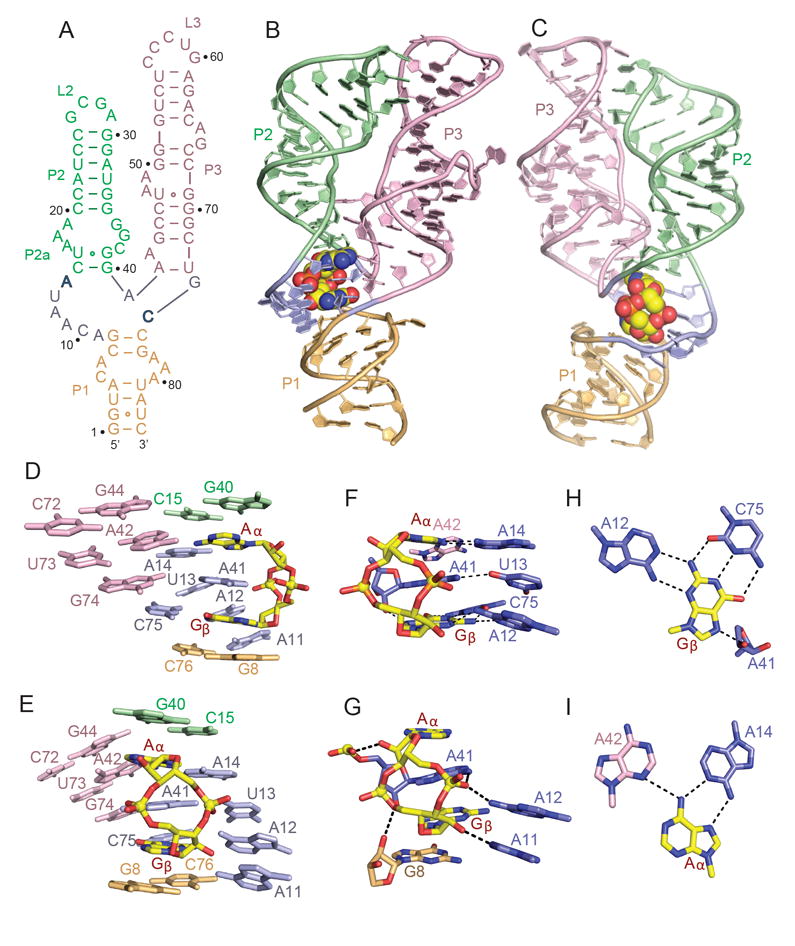
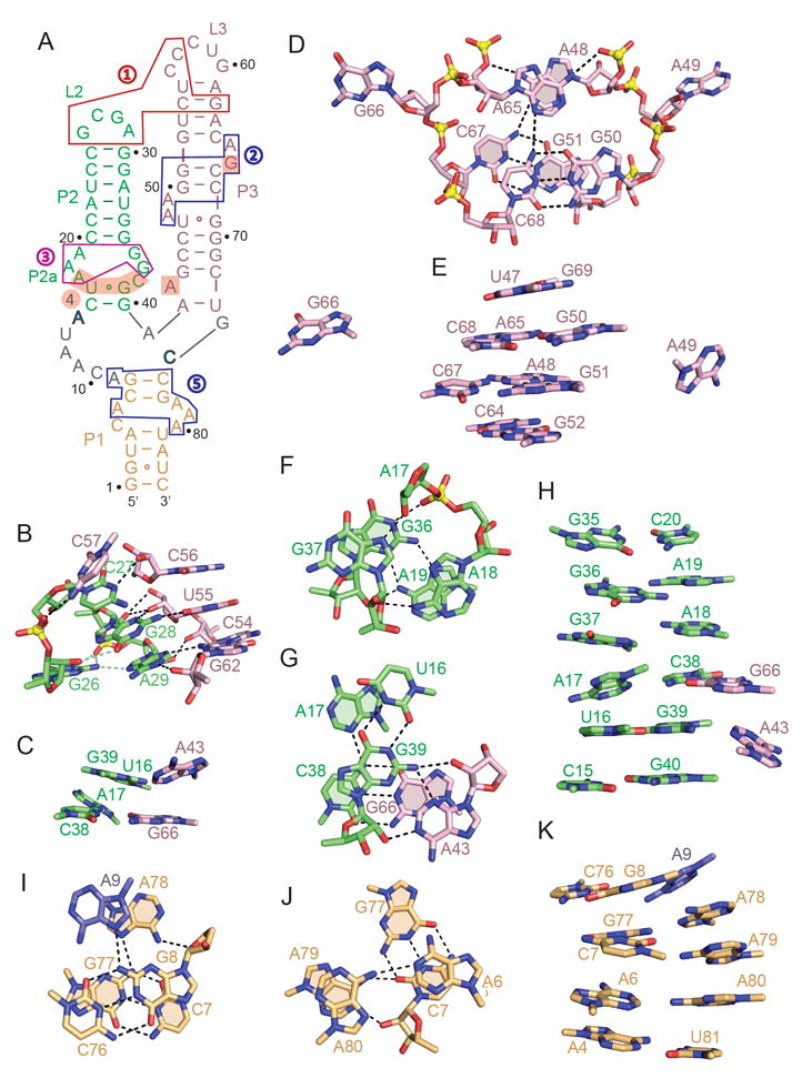
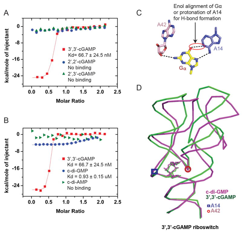
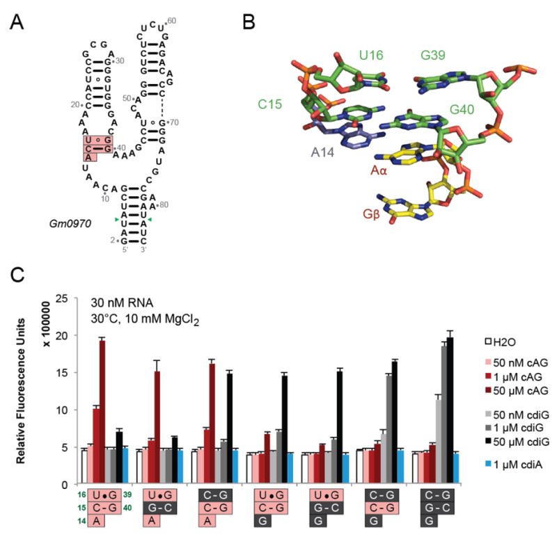
Cyclic dinucleotides are second messengers that target the adaptor STING and stimulate the innate immune response in mammals. Besides protein receptors, there are bacterial riboswitches that selectively recognize cyclic dinucleotides. We recently discovered a natural riboswitch that targets 3',3'-cGAMP, which is distinguished from the endogenous mammalian signal 2',3'-cGAMP by its backbone connectivity. Here, we report on structures of the aptamer domain of the 3',3'-cGAMP riboswitch from Geobacter in the 3',3'-cGAMP and c-di-GMP bound states. The riboswitch adopts a tuning fork-like architecture with a junctional ligand-binding pocket and different orientations of the arms are correlated with the identity of the bound cyclic dinucleotide. Subsequent biochemical experiments revealed that specificity of ligand recognition can be affected by point mutations outside of the binding pocket, which has implications for both the assignment and reengineering of riboswitches in this structural class.


Figures

Figure 1
Figure 1. Sequence, Structure and Pairing Alignments of the 3’,3’-cGAMP Riboswitch with bound 3’,3’-cGAMP
(A) Schematic of the secondary structure of the Gs1761 3’,3’-cGAMP riboswitch. This sequence differs from the natural riboswitch by containing A2G, U72C and C73U substitutions. (B, C) Two alternate views of the 2.05 A structure of the 3’,3’-cGAMP riboswitch with bound 3’,3’-cGAMP. The riboswitch RNA is shown in a ribbon representation and color coded by segments, while the bound 3’,3’-cGAMP is shown in a space-filling representation. Residues A14 and C74 that interact through base pairing with the bound cGAMP are shown in bold. (D, E) Two alternate views showing base stacking alignments within the 3’,3’-c-GAMP riboswitch (color-coded) centered about the bound 3’,3’-cGAMP (in yellow). (F, G) Two alternate views showing intermolecular base-base (panel F) and base-sugar-phosphate (panel G) hydrogen bonding interactions between 3’,3’-cGAMP (in yellow) and the riboswitch RNA residues (color-coded) centered about the binding site in the complex. (H) Intermolecular recognition of Gβ of 3’,3’-cGAMP (in yellow) by base-base hydrogen bonding with riboswitch RNA bases (in blue) in the complex. (I) Intermolecular recognition of Aα of 3’,3’-cGAMP (in yellow) by base-base hydrogen bonding with riboswitch RNA bases (color-coded) in the complex. See also Table S1 and Figure S1.
Figure 2
Figure 2. Long-range Loop-Loop Interactions and Topology of an Internal Bulge in Stem P3, P2, and P1 in the Complex
(A) Schematic of the secondary structure of the 3’,3’-cGAMP riboswitch highlighting loop-loop receptor interactions (boxed segment labeled 1 in red) and internal bulges in stem P3 (boxed segment labeled 2 in blue), stem P2 (boxed segments labeled 3 in magenta and labeled 4 with shaded background) and stem P1 (boxed segment labeled 5 in blue) in the complex. (B) Topology and intermolecular hydrogen bonding between residues in stem-loops P2 (in green) and P3 (in pink) in the complex represented by the boxed region (in red) labeled 1 in panel A. (C) Stacked triples between looped out bases from stem P3 (in pink) and the minor groove of base pairs in stem P2 (in green) that contribute to parallel alignment of stems P2 and P3 in the complex. (D) Topology and intermolecular hydrogen bonding between residues in the internal bubble in the complex represented by the boxed region (in blue) labeled 2 in panel A. (E) Stacking alignments spanning the bulged bases in stem P3 of the complex. (F) Topology and intermolecular hydrogen bonding between residues in the internal bulge in the complex represented by the boxed region (in magenta) labeled 3 in panel A. (G) Topology and intermolecular hydrogen bonding between residues in the internal bulge in the complex represented by the shaded region background labeled 4 in panel A. (H) Stacking alignments spanning the bulged bases in stem P2 of the complex. (I, J) Topology and intermolecular hydrogen bonding between residues in the internal bulge in stem P1 in the complex represented by the boxed region (in blue) labeled 5 in panel A. (K) Stacking alignments spanning the bulged bases in stem P1 of the complex. See also Table S1.
Figure 3
Figure 3. ITC-based Binding Curves of Cyclic Dinucleotides to the 3’,3’-cGAMP Riboswitch containing A2G, U72C and C73U substitutions
(A) ITC-based studies of the binding of 3’,3’-cGAMP, 2’,2’-cGAMP and 2’,2’-cGAMP to the 3’,3’-cGAMP riboswitch containing A2G, U72C and C73U substitutions. (B) ITC-based studies of the binding of 3’,3’-cGAMP, c-di-GMP and c-di-AMP to the 3’,3’-cGAMP riboswitch containing A2G, U72C and C73U substitutions. (C) Pairing of Gα with A14 in the structure of the complex of c-di-GMP with the 3’,3’-cGAMP riboswitch. (D) Superposition of the structures of the complexes of 3’,3’-cGAMP riboswitch with bound 3’,3’-cGAMP (in green) and c-di-GMP (in magenta). The positions of G42 and A14 are indicated by a closed circles and squares in the two complexes. See also Table S2.
Figure 4
Figure 4. A Single Mutation in the c-di-GMP Vc2 Riboswitch Facilitates Binding by 3’,3’-cGAM
(A) Sequence of the aptamer domain of the c-di-GMP Vc2 riboswitch (Smith et al. 2009). The color-coding is the same as in Figure 1A. (B) The Gα•G20 non-canonical pairing alignment in the structure of c-di-GMP bound to the c-di-GMP Vc2 riboswitch (PDB: 3IRW ; Smith et al. 2009). (C) Superposition of the structures of the c-di-GMP Vc2 riboswitch bound to c-di-GMP (in orange; PDB: 3IRW ; Smith et al. 2009) and G20A mutant c-di-GMP Vc2 riboswitch bound to 3’,3’-cGAMP (in blue). (D) The Aα•A20 non-canonical pairing alignment in the structure of 3’,3’-cGAMP bound to the c-di-GMP riboswitch. See also Table S3
Figure 5
Figure 5. The P2a Region Affects Ligand Selectivity of the Gm0970 3’,3’-cGAMP Riboswitch
(A) Secondary structure of Gm0970, another 3’,3’-cGAMP-selective riboswitch. The nucleotide numbering is set to match that of Gs1761 in the P2a region (pink boxes). Green arrows indicate the positions in the P1 stem to which the Spinach aptamer was fused. (B) Structure of the P2a region for Gs1761 with bound 3’,3’-cGAMP. (C) Spinach-based selectivity screen of Gm0970 riboswitch constructs with mutations to the P2a region shown. Fluorescence activation was measured in the presence of no ligand or different cyclic dinucleotides at the indicated concentrations. The nucleotides from Gm0970 (pink) were changed to those from the c-di-GMP-selective riboswitch, Vc2 (gray).

References

    1. Ablasser A, Goldeck M, Cavlar T, Deimling T, Witte G, Rohl I, Hopfner KP, Ludwig J, Hornung V. cGAS produces a 2’-5’-linked cyclic dinucleotide second messenger that activates STING. Nature. 2013;498:380–384. - PMC - PubMed
    1. Adams PD, Grosse-Kunstleve RW, Hung LW, Loerger TR, McCoy AJ, Moriarty NW, Read RJ, Sacchettini JC, Sauter NK, Terwilliger TC. PHENIX: building new software for automated crystallographic structure determination. Acta Crystallogr D Biol Crystallogr. 2002:1948–1954. - PubMed
    1. Batey RT, Gilbert SD, Montange RK. Structure of a natural guanine-responsive riboswitch complexed with the metabolite hypoxanthine. Nature. 2004;432:411–415. - PubMed
    1. Burdette DL, Monroe KM, Sotelo-Troha K, Iwig JS, Eckert B, Hyodo M, Hayakawa Y, Vance RE. STING is a direct innate immune sensor of cyclic di-GMP. Nature. 2011;478:515–518. - PMC - PubMed
    1. Cai X, Chiu YH, Chen ZJ. The cGAS-cGAMP-STING pathway of cytosolic DNA sensing and signaling. Mol Cell. 2014;54:289–296. - PubMed

Publication types

Associated data