. 2015 Apr 7;11(1):61-70.
doi: 10.1016/j.celrep.2015.03.003. Epub 2015 Mar 26.

Structure and function of Neisseria gonorrhoeae MtrF illuminates a class of antimetabolite efflux pumps

Affiliations

Structure and function of Neisseria gonorrhoeae MtrF illuminates a class of antimetabolite efflux pumps

Chih-Chia Su et al. Cell Rep. .

Abstract

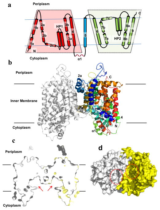
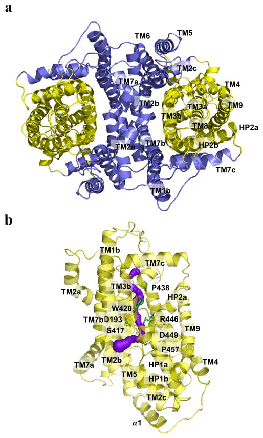
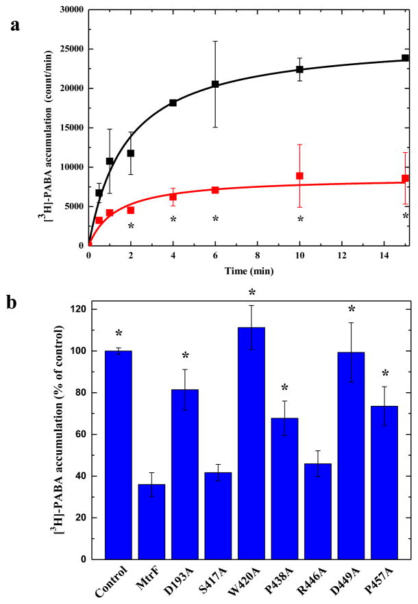
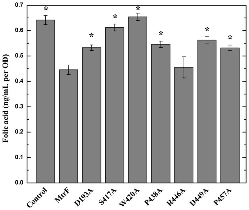
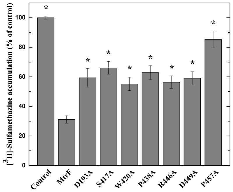
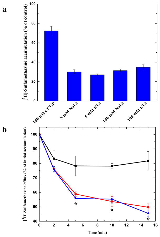
Neisseria gonorrhoeae is an obligate human pathogen and the causative agent of the sexually transmitted disease gonorrhea. The control of this disease has been compromised by the increasing proportion of infections due to antibiotic-resistant strains, which are growing at an alarming rate. N. gonorrhoeae MtrF is an integral membrane protein that belongs to the AbgT family of transporters for which no structural information is available. Here, we describe the crystal structure of MtrF, revealing a dimeric molecule with architecture distinct from all other families of transporters. MtrF is a bowl-shaped dimer with a solvent-filled basin extending from the cytoplasm to halfway across the membrane bilayer. Each subunit of the transporter contains nine transmembrane helices and two hairpins, posing a plausible pathway for substrate transport. A combination of the crystal structure and biochemical functional assays suggests that MtrF is an antibiotic efflux pump mediating bacterial resistance to sulfonamide antimetabolite drugs.


Figures

Fig. 1
Fig. 1
Structure of the N. gonorrhoeae MtrF transporter. (a) Transmembrane topology of N. gonorrhoeae MtrF. The transporter contains nine transmembrane helices (TMs) and two hairpins (HPs). (b) Ribbon diagram of a dimer of MtrF viewed in the membrane plane. The right subunit of the dimer is colored using a rainbow gradient from the N-terminus (blue) to the C-terminus (red), whereas the left subunit is colored gray. The MtrF dimer forms a bowl-shaped structure with a concave aqueous basin facing the intracellular solution. (c) Surface representation of a cross section of the MtrF dimer sliced through the middle of the protein. Each protomer forms an internal cavity (red arrow), which is accessible to the cytoplasm. (d) Bottom view of a surface representation of the MtrF dimer, indicating a solvent accessible cavity (red circle) from each protomer of the protein. The two protomers are colored gray and yellow.
Fig. 2
Fig. 2
Inner and Outer cores of MtrF. (a) The inner core of MtrF, comprising TMs 1, 2, 5, 6 and 7 (colored slate), contributes to dimerization as well as formation of a frame-like structure housing the outer core of the protomer. The outer core of MtrF is composed of TMs 3, 4, 8, 9 as well as HPs 1 and 2 (colored yellow). (b) The outer core of MtrF forms a channel (colored purple) spanning approximately from the middle of the inner membrane up to the periplasmic space. This channel was calculated using the program CAVER (http://loschmidt.chemi.muni.cz/caver). The secondary structural elements of the MtrF protomer are in yellow. Residues D193, S417, W420, P438, R446, D449, and P457 are in green sticks.
Fig. 3
Fig. 3
Accumulation of radioactive p-aminobenzoic acid. (a) Time course of [3H]-PABA accumulation by E. coli BL21(DE3)ΔabgTΔpabA double knockout cells transformed with pET15bΩmtrF or pET15b. Cells expressing mtrF (red curve) show a significant decrease in [3H]-PABA accumulation when compared with cells carrying the empty vector (black curve). “*” indicates values of BL21(DE3)ΔabgTΔpabA/pET15bΩmtrF cells that are significantly different from the control (BL21(DE3)ΔabgTΔpabA/pET15b) values (P < 0.05). (b) Mutants of the MtrF transporter. Cells possessing the mutant transporter D193A, W420A, P438A, D449A and P457A show a significant increase in the level of [3H]-PABA accumulations compared with cells expressing wild-type MtrF. However, cells expressing S417A and R446A only show a modest change on the [3H]-PABA concentration when compared with cells carrying wild-type MtrF. “*” indicates values of BL21(DE3)ΔabgTΔpabA/pET15b and BL21(DE3)ΔabgTΔpabA cells expressing the mutant transporters that are significantly higher than that of BL21(DE3)ΔabgTΔpabA/pET15bΩmtrF expressing wild-type MtrF (P < 0.009). The data showed in (a) and (b) are the cumulative average of three successive recordings.
Fig. 4
Fig. 4
Intracellular folic acid concentration. Folic acid concentration in E. coli BL21(DE3)ΔabgTΔpabA double knockout cells expressing MtrF were markedly reduced in comparison with cells transformed with the empty vector. When transformed with plasmid expressing the mutant transporter, D193A, S417A, W420A, P438A, D449A, and P457A, folic acid production was significantly increased in these cells. However, the level of intracellular folic acid concentration in BL21(DE3)ΔabgTΔpabA cells expressing R446A was nearly identical to that of the double knockout strain carrying wild-type MtrF. Each bar represents the mean of three separate cultures. “*” indicates values of BL21(DE3)ΔabgTΔpabA/pET15b and BL21(DE3)ΔabgTΔpabA cells expressing the mutant transporters that are significantly higher than that of BL21(DE3)ΔabgTΔpabA/pET15bΩmtrF expressing wild-type MtrF (P < 0.04).
Fig. 5
Fig. 5
Accumulation of radioactive sulfamethazine. E. coli BL21(DE3)ΔabgTΔpabA cells expressing MtrF show a significant decrease in [3H]-sulfamethazine accumulation when compared with cells carrying the empty vector. When transformed with plasmids expressing the mutant transporters, D193A, S417A, W420A, P438A, R446A, D449A, and P457A, the levels of intracellular [3H]-sulfamethazine accumulation were much higher than that of cells expressing wild-type MtrF. Each bar represents the mean of three different cultures. “*” indicates values of BL21(DE3)ΔabgTΔpabA/pET15b and BL21(DE3)ΔabgTΔpabA cells expressing the mutant transporters that are significantly higher than that of BL21(DE3)ΔabgTΔpabA/pET15bΩmtrF expressing wild-type MtrF (P < 0.001).
Fig. 6
Fig. 6
Transport of sulfamethazine via MtrF. (a) Accumulation of radioactive sulfamethazine in BL21(DE3)ΔabgTΔpabA/pET15bΩmtrF cells with different sodium or potassium ion concentrations. The data indicate that the transport function of MtrF is independent of sodium or potassium ions. (b) Efflux of radioactive sulfamethazine in BL21(DE3)ΔabgTΔpabA/pET15bΩmtrF cells in the absence and presence of sodium ions. The presence of Na+ does not affect [3H]-sulfamethazine efflux in BL21(DE3)ΔabgTΔpabA/pET15bΩmtrF cells (black, controlled cells transformed with empty vector; red, 0 mM NaCl; blue, 5 mM NaCl). “*” indicates values of radioactive counts of intracellular [3H]-sulfamethazine in BL21(DE3)ΔabgTΔpabA/pET15bΩmtrF cells with 0 mM NaCl (red) (P < 0.003) and 5 mM NaCl (P < 0.001) that are significantly different from those of the control (black). The data showed in (a) and (b) are the cumulative average of three successive recordings.

References

    1. Adams PD, Grosse-Kunstleve RW, Hung LW, Ioerger TR, McCroy AJ, Moriarty NW, et al. PHENIX: building new software for automated crystallographic structure determination. Acta Crystallogr. 2002;58:1948–1954. - PubMed
    1. Bolla JR, Su CC, Do SV, Radhakrishnan A, Kumar N, Long F, Chou TH, Delmar JA, Lei HT, Rajashankar KR, Shafer WM, Yu EW. Crystal structure of the Neisseria gonorrhoeae MtrD inner membrane multidrug efflux pump. PLoS One. 2014;9:e97903. - PMC - PubMed
    1. Brown MH, Paulsen IT, Skurray RA. The multidrug efflux protein NorM is a prototype of a new family of transporters. Mol Microbiol. 1999;31:394–395. - PubMed
    1. Brünger AT, Adams PD, Clore GM, DeLano WL, Gros P, Grosse-Kunstleve RW, Jiang JS, Kuszewski J, Nilges M, Pannu NS, Read RJ, Rice LM, Simonson T, Warren GL. Crystallography & NMR system: A new software suite for macromolecular structure determination. Acta Crystallogr. 1998;D54:905–921. - PubMed
    1. Carter EL, Jager L, Gardner L, Hall CC, Willis S, Green JM. Escherichia coli abg genes enable uptake and cleavage of the folate catabolite p-aminobenzoyl-glutamate. J Bacteriol. 2007;189:3329–3334. - PMC - PubMed

Publication types

MeSH terms

Associated data

LinkOut - more resources