

ADRA2A is involved in neuro-endocrine regulation of bone resorption
- PMID: 25818344
- PMCID: PMC4511350
- DOI: 10.1111/jcmm.12505
ADRA2A is involved in neuro-endocrine regulation of bone resorption
Abstract
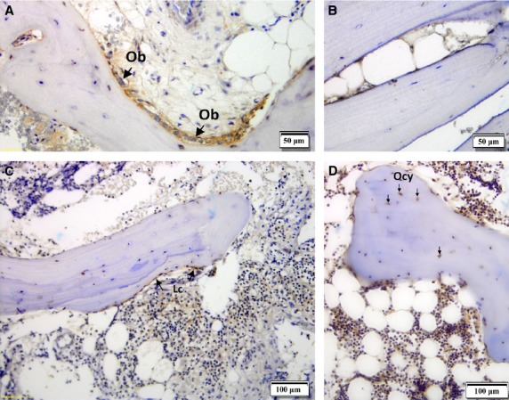
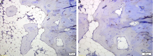
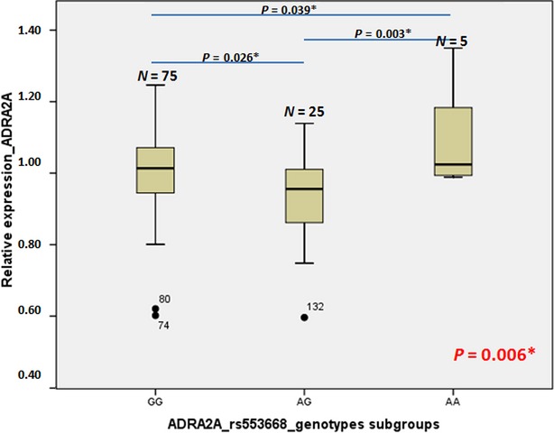
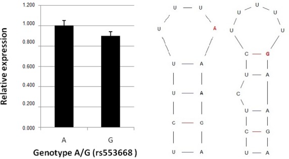
Adrenergic stimulation is important for osteoclast differentiation and bone resorption. Previous research shows that this happens through β2-adrenergic receptor (AR), but there are conflicting evidence on presence and role of α2A-AR in bone. The aim of this study was to investigate the presence of α2A-AR and its involvement in neuro-endocrine signalling of bone remodelling in humans. Real-time polymerase chain reaction (PCR) and immunohistochemistry were used to investigate α2A-AR receptor presence and localization in bone cells. Functionality of rs553668 and rs1800544 single nucleotide polymorphism SNPs located in α2A-AR gene was analysed by qPCR expression on bone samples and luciferase reporter assay in human osteosarcoma HOS cells. Using real-time PCR, genetic association study between rs553668 A>G and rs1800544 C>G SNPs and major bone markers was performed on 661 Slovenian patients with osteoporosis. α2A-AR is expressed in osteoblasts and lining cells but not in osteocytes. SNP rs553668 has a significant influence on α2A-AR mRNA level in human bone samples through the stability of mRNA. α2A-AR gene locus associates with important bone remodelling markers (BMD, CTX, Cathepsin K and pOC). The results of this study are providing comprehensive new evidence that α2A-AR is involved in neuro-endocrine signalling of bone turnover and development of osteoporosis. As shown by our results the neurological signalling is mediated through osteoblasts and result in bone resorption. Genetic study showed association of SNPs in α2A-AR gene locus with bone remodelling markers, identifying the individuals with higher risk of development of osteoporosis.
Keywords: ADRA2A; adrenergic signalling; bone; osteoblast; osteoporosis; resorption.
© 2015 The Authors. Journal of Cellular and Molecular Medicine published by John Wiley & Sons Ltd and Foundation for Cellular and Molecular Medicine.
Figures
References
-
- Ralston SH, de Crombrugghe B. Genetic regulation of bone mass and susceptibility to osteoporosis. Genes Dev. 2006;20:2492–506. - PubMed
-
- Trost Z, Trebse R, Prezelj J, et al. A microarray based identification of osteoporosis-related genes in primary culture of human osteoblasts. Bone. 2010;46:72–80. - PubMed
-
- Takeuchi T, Tsuboi T, Arai M, et al. Adrenergic stimulation of osteoclastogenesis mediated by expression of osteoclast differentiation factor in MC3T3-E1 osteoblast-like cells. Biochem Pharmacol. 2001;61:579–86. - PubMed
-
- Ducy P, Amling M, Takeda S, et al. Leptin inhibits bone formation through a hypothalamic relay: a central control of bone mass. Cell. 2000;100:197–207. - PubMed
Publication types
MeSH terms
Substances
LinkOut - more resources
Full Text Sources
Other Literature Sources
Molecular Biology Databases
Research Materials
