Review
. 2015 Mar 27;7(5):a020388.
doi: 10.1101/cshperspect.a020388.

Astrocyte regulation of blood flow in the brain

Affiliations
Review

Astrocyte regulation of blood flow in the brain

Brian A MacVicar et al. Cold Spring Harb Perspect Biol. .

Abstract

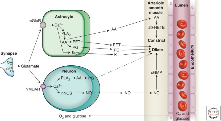
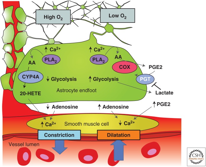
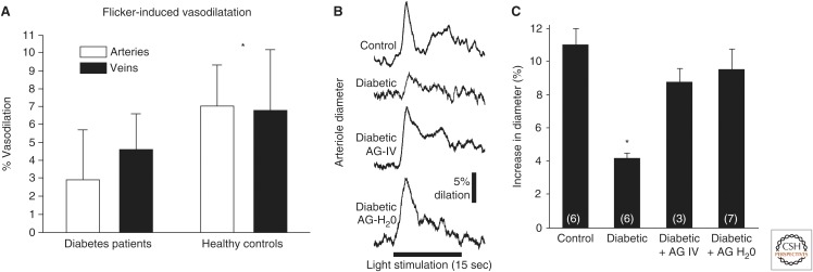
Neuronal activity results in increased blood flow in the brain, a response named functional hyperemia. Astrocytes play an important role in mediating this response. Neurotransmitters released from active neurons evoke Ca(2+) increases in astrocytes, leading to the release of vasoactive metabolites of arachidonic acid from astrocyte endfeet onto blood vessels. Synthesis of prostaglandin E2 (PGE2) and epoxyeicosatrienoic acids (EETs) dilate blood vessels, whereas 20-hydroxyeicosatetraenoic acid (20-HETE) constricts vessels. The release of K(+) from astrocyte endfeet may also contribute to vasodilation. Oxygen modulates astrocyte regulation of blood flow. Under normoxic conditions, astrocytic Ca(2+) signaling results in vasodilation, whereas under hyperoxic conditions, vasoconstriction is favored. Astrocytes also contribute to the generation of vascular tone. Tonic release of both 20-HETE and ATP from astrocytes constricts vascular smooth muscle cells, generating vessel tone. Under pathological conditions, including Alzheimer's disease and diabetic retinopathy, disruption of normal astrocyte physiology can compromise the regulation of blood flow.


Figures

Figure 1.
Figure 1.
Drawing of brain astrocytes by Santiago Ramón y Cajal. (A,B) Astrocytes, the darker cells in the drawing, contact both neurons, (C,D) the lighter cells, and (F) a blood vessel. As suggested by Ramón y Cajal, astrocytes are ideally situated to mediate signaling from neurons to blood vessels and to increase cerebral blood flow (CBF) in response to neuronal activity.
Figure 2.
Figure 2.
Summary of signaling pathways that mediate neurovascular coupling in the brain. Synaptically released glutamate acts on N-methyl-d-aspartate receptors (NMDARs) in neurons to increase [Ca2+]i, causing neuronal nitric oxide synthase (nNOS) to release nitric oxide (NO), which activates smooth muscle guanylate cyclase. Raised [Ca2+]i may also (dashed line) generate arachidonic acid (AA) from phospholipase A2 (PLA2), which is converted to prostaglandins (PG) that dilate vessels. Glutamate also raises [Ca2+]i in astrocytes by activating metabotropic glutamate receptors (mGluR), generating arachidonic acid, and three types of AA metabolites: prostaglandins and EETs in astrocytes, which dilate vessels, and 20-HETE in smooth muscle, which constricts vessels. An increase of [Ca2+]i in astrocyte endfeet may also activate Ca2+-gated K+ channels (gk(Ca), alternative abbreviation, BK), releasing K+, which dilates vessels. (From Attwell et al. 2010; reprinted, with permission, from the authors.)
Figure 3.
Figure 3.
Summary of the modulation of arachidonic acid metabolite-mediated neurovascular coupling by tissue pO2 and lactate. Intracellular Ca2+ transients activate PLA2 to liberate arachidonic acid (AA) from plasma membrane lipids. In high pO2, AA is converted to 20-HETE, which increases intracellular Ca2+ in smooth muscle cells, causing vasoconstriction (left side). Extracellular adenosine that can inhibit Ca2+ entry into smooth muscle cells is also lower in high tissue pO2, further promoting vasoconstriction. In lower pO2 (right side), the vasodilation pathway dominates and AA is converted to PGE2 by COX-1 and is released by diffusion. Extracellular PGE2 is cleared from the extracellular space by the prostaglandin transporter (PGT), which is expressed in astrocytes and neurons. When pO2 decreases, glycolysis is enhanced and extracellular levels of lactate increase. Extracellular lactate attenuates PGE2 uptake by PGT leading to higher extracellular PGE2 and enhanced relaxation of smooth muscle tone and vasodilation. In addition, higher levels of extracellular adenosine reduces smooth muscle cell constriction via A2A activation. (From Gordon et al. 2011; reprinted, with permission, from the authors.)
Figure 4.
Figure 4.
Flicker-induced vasodilation of retinal blood vessels is depressed in diabetic retinopathy. (A) Flicker-induced vasodilation of primary arterioles and venules is depressed in patients with type 1 diabetes. (Panel A is from Pemp et al. 2009; reprinted, with permission, from the Association for Research in Vision and Ophthalmology © 2009.) B and C show that the depression of flicker-induced vasodilation in diabetic rats is reversed by the iNOS inhibitor aminoguanidine (AG). (B) Both acute AG administration (AG-IV) and chronic administration in drinking water (AG-H2O) reverses the loss of flicker-induced vasodilation in diabetic animals in vivo. (C) Summary of results. (Panels B and C are from Mishra and Newman 2012; reprinted, with permission, from the authors and Frontiers in Neuroscience Creative Common attribution license.)

References

    1. Akiyama H, Barger S, Barnum S, Bradt B, Bauer J, Cole GM, Cooper NR, Eikelenboom P, Emmerling M, Fiebich BL, et al. 2000. Inflammation and Alzheimer’s disease. Neurobiol Aging 21: 383–421. - PMC - PubMed
    1. Alzheimer A. 1907. Über eine eigenartige Erkrankung der Hirnrinde [About a peculiar disease of the cerebral cortex]. Allg Z Psychiat 64: 146–148.
    1. Attwell D, Buchan AM, Charpak S, Lauritzen M, MacVicar BA, Newman EA. 2010. Glial and neuronal control of brain blood flow. Nature 468: 232–243. - PMC - PubMed
    1. Bekar LK, Wei HS, Nedergaard M. 2012. The locus coeruleus-norepinephrine network optimizes coupling of cerebral blood volume with oxygen demand. J Cereb Blood Flow Metab 32: 2135–2145. - PMC - PubMed
    1. Blinder P, Tsai PS, Kaufhold JP, Knutsen PM, Suhl H, Kleinfeld D. 2013. The cortical angiome: An interconnected vascular network with noncolumnar patterns of blood flow. Nat Neurosci 16: 889–897. - PMC - PubMed

Publication types

LinkOut - more resources