. 2015 Mar 30:6:6641.
doi: 10.1038/ncomms7641.

Skp2-macroH2A1-CDK8 axis orchestrates G2/M transition and tumorigenesis

Affiliations

Skp2-macroH2A1-CDK8 axis orchestrates G2/M transition and tumorigenesis

Dazhi Xu et al. Nat Commun. .

Abstract

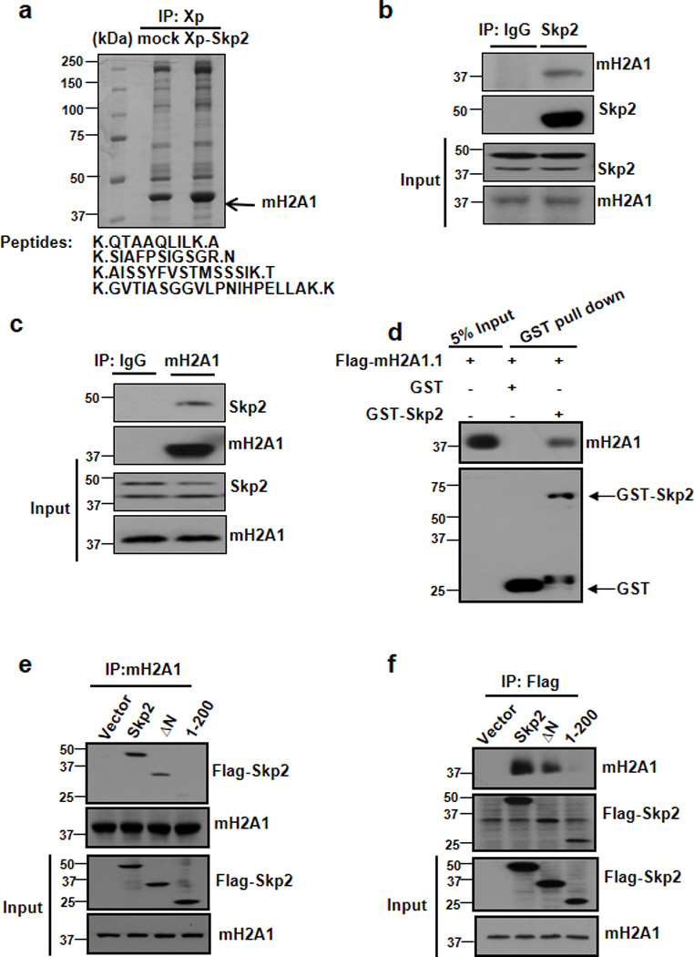
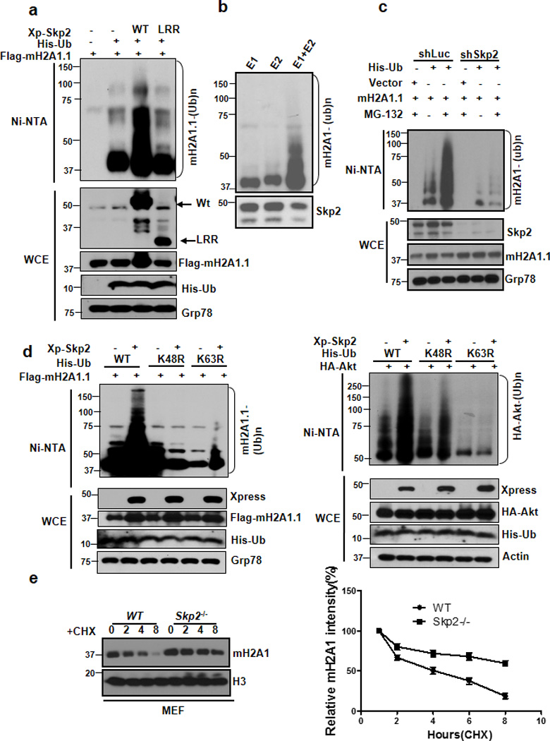
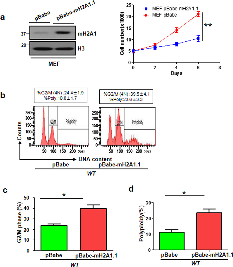
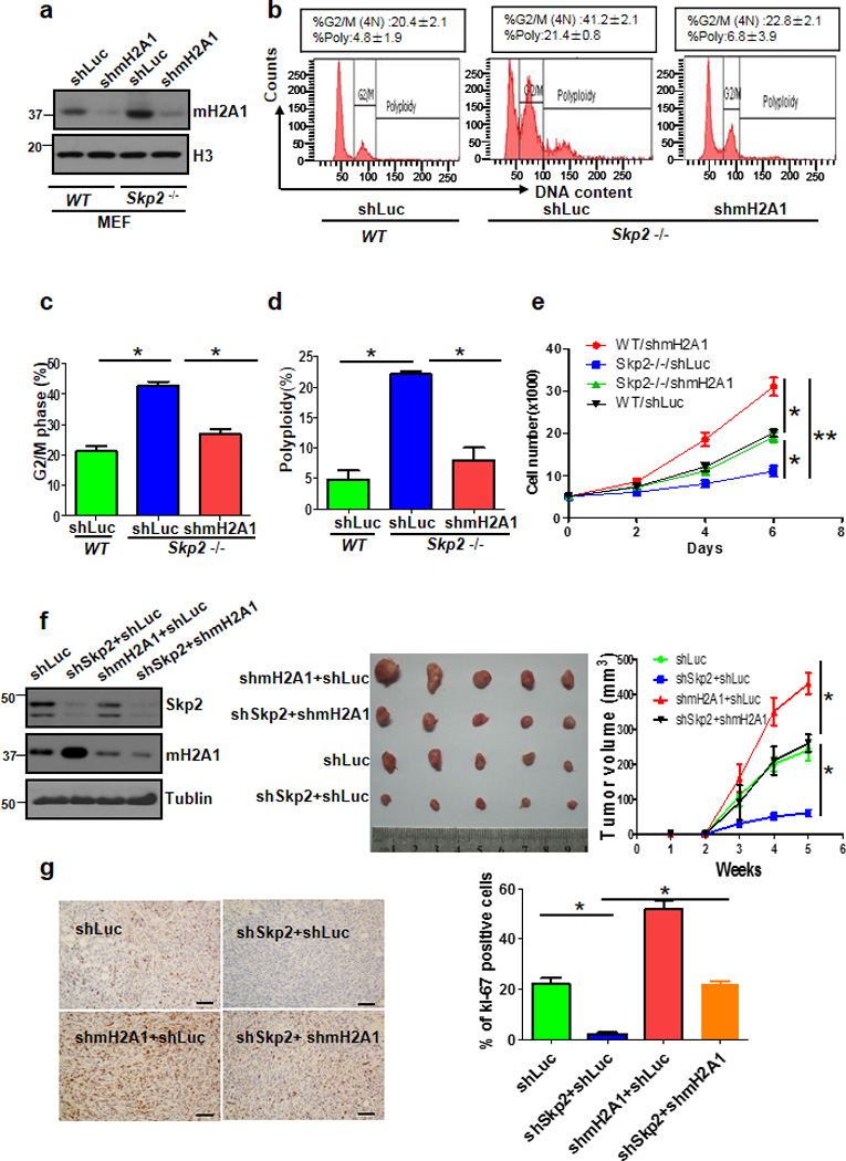
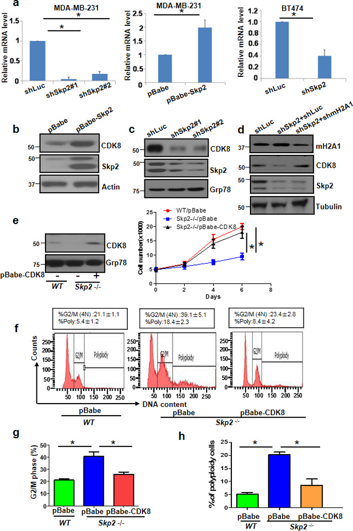
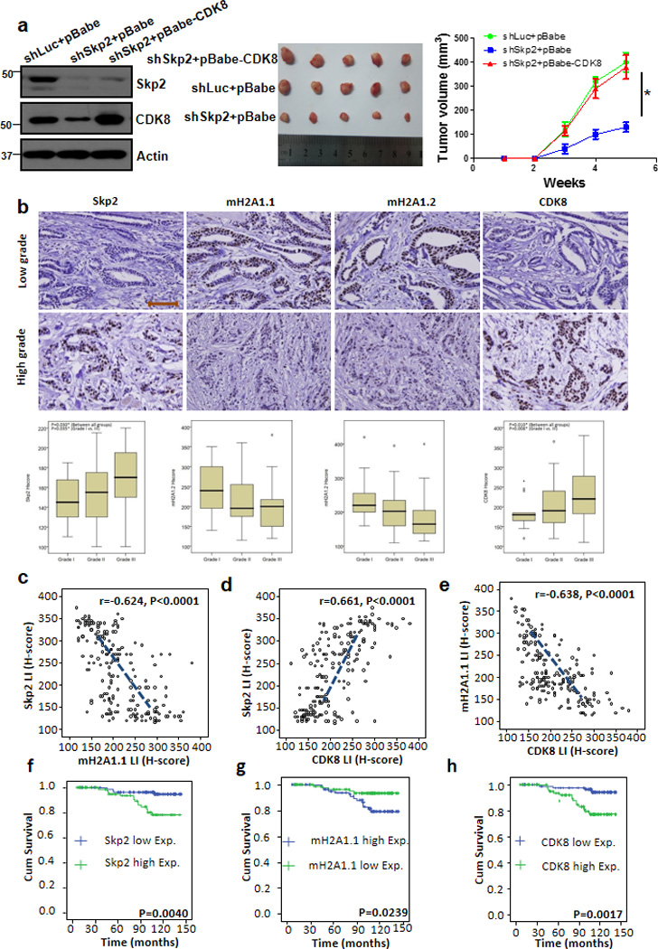
Understanding the mechanism by which cell growth, migration, polyploidy, and tumorigenesis are regulated may provide important therapeutic strategies for cancer therapy. Here we identify the Skp2-macroH2A1 (mH2A1)-cyclin-dependent kinase 8 (CDK8) axis as a critical pathway for these processes, and deregulation of this pathway is associated with human breast cancer progression and patient survival outcome. We showed that mH2A1 is a new substrate of Skp2 SCF complex whose degradation by Skp2 promotes CDK8 gene and protein expression. Strikingly, breast tumour suppression on Skp2 deficiency can be rescued by mH2A1 knockdown or CDK8 restoration using mouse tumour models. We further show that CDK8 regulates p27 protein expression by facilitating Skp2-mediated p27 ubiquitination and degradation. Our study establishes a critical role of Skp2-mH2A1-CDK8 axis in breast cancer development and targeting this pathway offers a promising strategy for breast cancer therapy.


Figures

Figure 1
Figure 1. Skp2 interacts with mH2A1
(a) Lysates from 293T stably transfected with vector or Xpress-Skp2 (XP-Skp2) were immunopreicipated with XP antibody and subjected to mass spectrometry analysis. Histone variant mH2A1 was identified as a novel binding partner for Skp2. (b, c) 293T cells were harvested for immunoprecipitation with Skp2 antibody (b) or mH2A1 antibody (c), followed by immunoblotting. (d) In vitro GST pull down assay using recombinant GST, GST-Skp2 proteins purified from bacteria and in vitro translated Flag-mH2A1.1. 5% of input was loaded in this assay. (e, f) 293T cells were transfected with indicated plasmids and harvested for immunoprecipitation assay.
Figure 2
Figure 2. Skp2 SCF is a direct E3 ligase for mH2A1 and triggers ubiquitination and degradation of mH2A1
(a) In vivo ubiquitination assay from 293T cells transfected with indicated plasmids. Ni-NTA: nickel bead pulldown. WCE, whole-cell extracts. (b) Flag-Skp2 proteins isolated 293T cells transfected with Flag-Skp2 were incubated with ATP, E1, E2 along with mH2A1 proteins isolated from 293T cells for in vitro ubiquitination assay. (c) 293T cells stably expressing luciferase shRNA (shLuc) or Skp2 shRNA (shSkp2) were transfected with the indicated plasmids and harvested for in vivo ubiquitination assay. (d) In vivo ubiquitination assay of 293T cells transfected with His-Ubiquitin (His-Ub), His-Ub K48R, or His-Ub K63R along with other constructs. (e) Primary WT and Skp2−/− MEFs were treated with cycloheximide for the indicated times and harvested for immunoblotting analysis. A representative blot and quantification from there independent experiments were shown. Cycloheximide: CHX.
Figure 3
Figure 3. mH2A1.1 overexpression in primary MEFs triggers cell growth arrest and polyploidy
(a) Primary MEFs were infected with pBabe or pBabe-mH2A1.1 lentiviral RNAs, selected, and harvested for Immunoblotting (Left panel). These cells were plated in 24-well plated for cell growth assay using direct cell counting (Right panel). (b) Flow cytometry analysis of DNA content in primary MEFs stably expressing pBabe or pBabe-mH2A1.1. (c) G2/M phase was determined by Flow cytometry analysis of primary WT MEFs with stably expressing pBabe or pBabe-mH2A1.1. The quantified results are presented as means ± s.d. (d) Polyploidy of primary WT and Skp2−/− MEFs with stably expressing pBabe or pBabe-mH2A1.1. The quantified results are presented as means ± s.d. (Error bars indicate s.e.m. Data represent mean values of three independent experiments. Student’s t-test used; *p<0.05;**p<0.01)
Figure 4
Figure 4. mH2A1 deficiency rescues cell growth arrest and polyploidy upon Skp2 loss
(a) Primary WT and Skp2−/− MEFs were infected with lucifcerse shRNAs (shLuc) or mH2A1 shRNAs (shmH2A1), selected and harvested for Immunoblotting. (b) Flow cytometry analysis of DNA content in WT and Skp2−/− MEFs with Luc or mH2A1 knockdown. (c) G2/M phase was determined by Flow cytometry analysis of primary WT and Skp2−/− MEFs with stably expressing shLuc or shmH2A1. (d) Polyploidy of primary WT and Skp2−/− MEFs with stably expressing shLuc or shmH2A1. (e) Cell growth assay by direct cell counting of WT and Skp2−/− MEFs with Luc or mH2A1 knockdown. (f) MDA-MB-231 cells infected with various shRNAs were subcutaneously injected into nude mice (n=5 for each group). Tumour size was monitored and calculated by caliper for up to 5 weeks (see Methods). The cell lysates from tumour cells were subjected to immunoblotting (left panel). A photo of five tumours aligned together were presented (middle panel). The results were calculated as mean values ± s.d. (g)MDA-MB-231 cells infected with various shRNAs were subcutaneously injected into nude mice, and breast tumours were harvested from nude mice at week 5 for Ki-67 staining by IHC and quantitated (Scale bars, 50µm) (Error bars indicate s.e.m. Data represent mean values of three independent experiments. Student’s t-test used; *p<0.05;**p<0.01)
Figure 5
Figure 5. CDK8 is a downstream effector responsible for Skp2-mediated cell growth, G2/M and polyploidy
(a) The mRNA levels of CDK8 were measured by real-time PCR in MDA-MB-231 and BT474 cells with Luc or Skp2 knockdown (n=3). (b-d) Immunoblotting of MDA-MB-231 cells infected with pBabe or pBabe-Skp2 (B), infected with Luc or Skp2 shRNAs (c) or infected with Luc shRNAs, Skp2 shRNAs, or Skp2 plus mH2A1 shRNAs (d). (e) Immunoblotting assay (Left panel) and cell growth assay by direct cell counting (Right panel) of primary WT and Skp2−/− MEFs with stably expressing pBabe or pBabe-CDK8. (f) Flow cytometry analysis of DNA content in primary WT and Skp2−/− MEFs with stably expressing pBabe or pBabe-CDK8. (g) G2/M phase was determined by Flow cytometry analysis of primary WT MEFs with stably expressing pBabe or pBabe-CDK8. (h) Polyploidy of primary WT and Skp2−/− MEFs with stably expressing pBabe or pBabe-CDK8. (Error bars indicate s.e.m. Data represent mean values of three independent experiments. Student’s t-test used; *p<0.05)
Figure 6
Figure 6. CDK8 restoration recues the defect in tumourigenesis upon Skp2 loss, and Skp2 expression is inversely correlated with mH2A1, but positively correlated with CDK8 expression in human breast cancer samples
(a) Tumour volume (mm3) post-subcutaneous injections of MDA-MB-231 cells; *P < 0.05 (n=5 mice per group). MDA-MB-231 cells silenced with control, Skp2 shRNAs, or Skp2 shRNAs plus CDK8 overexpression were injected into nude mice (n=5 for each group) and followed up for tumourigenesis (see Methods). The lysate of tumour cells were subjected to immunoblotting (left panel). A photo of five tumours aligned together were presented (middle panel). The results were calculated as mean values ± s.d. *p<0.05, **p<0.01 using Student’s t-test (right panel). (b) Histological analysis of Skp2, mH2A1.1, mH2A1.2 and CDK8 expressions in patients with low or high grade of breast invasive ductal carcinoma. Scale bar represents 200µm. (c-e) Skp2 expression was negatively correlated with mH2A1.1 (r=−0.624, p<0.001) (c), but positively associated with CDK8 (r=0.661, p<0.001) (d). mH2A1.1 negatively correlated with CDK8 expression (r=−0.638, p<0.001) (e). (f-h), Kaplan-Meier plot analysis of overall survival of 189 cases of breast invasive ductal carcinoma patients with low or high expression of Skp2 (f), mH2A1.1 (g) or CDK8 (h) P-value in all case is < 0.01 by using Mann-Whitney U test.
Figure 7
Figure 7. CDK8 phosphorylate p27 at T187 and facilitates Skp2-mediated p27 ubiquitination and degradation
(a) 293T cells stably expressing luciferase shRNA (shLuc) or Skp2 shRNA (shSkp2) were transfected with the indicated plasmids and harvested for in vivo ubiquitination assay. (b) 293T cells stably expressing luciferase shRNA (shLuc) or CDK8 shRNA (shCDK8) were transfected with the indicated plasmids and harvested for in vivo ubiquitination assay. (c) 293T cells stably expressing luciferase shRNA (shLuc) or CDK8 shRNA (shCDK8) were transfected with the indicated plasmids and harvested for in vivo ubiquitination assay. (d) Cell lysates from 293T cells transfected with vector or CDK8 along with WT Flag-p27 or Flag-p27-187A were immunoprecipitated with Flag antibody and subjected to the immunoblotting assay. p27 phosphorylation at T187 was detected by using an antibody against phosphorylated p27 at T187. (e) Cell lysates from Luc or CDK8 knockdown 293T cells transfected with vector or Flag-p27 were immunoprecipitated with Flag antibody and subjected to the immunoblotting assay. p27 phosphorylation at T187 was detected by using an antibody against phosphorylated p27 at T187. (f) CDK8 phosphorylates p27 in vitro. Immunopurified Flag-p27 proteins were incubated with or without recombinant active CDK8 or CDK8-KD proteins and subjected to the immunoblotting assay. p27 phosphorylation at T187 was detected by using an antibody against phosphorylated p27 at T187. (g) In vivo ubiquitination assay of 293T cells transfected with Flag-p27 WT or Flag-p27 T187A along with other constructs. (h) The working model for Skp2 E3 ligase-mediated cell growth, cell migration, cell cycle checkpoint, polyploidy and tumourigenesis. Skp2 interacts with mH2A1 and induces its ubiquitination and degradation. mH2A1 degradation induced by Skp2 leads to gene and protein expression of CDK8, which may serve as a kinase for p27 or regulate CyclinA/CDK2 to drive p27 phosphorylation at T187, leading to promoting Skp2-mediated p27 ubiquitination and degradation, which may contribute to cell growth, cell migration, G2/M arrest, polyploidy and tumourigenesis. Alternatively, Skp2-mH2A1-CDK8 axis may regulate these processes independently of p27 regulation.

References

    1. Bakhoum SF, Compton DA. Chromosomal instability and cancer: a complex relationship with therapeutic potential. J Clin Invest. 2012;122:1138–1143. - PMC - PubMed
    1. Ribeiro PS, et al. Combined functional genomic and proteomic approaches identify a PP2A complex as a negative regulator of Hippo signaling. Mol Cell. 2010;39:521–534. - PubMed
    1. Talos F, Nemajerova A, Flores ER, Petrenko O, Moll UM. p73 suppresses polyploidy and aneuploidy in the absence of functional p53. Mol Cell. 2007;27:647–659. - PubMed
    1. Chan CH, et al. The Skp2-SCF E3 ligase regulates Akt ubiquitination, glycolysis, herceptin sensitivity, and tumorigenesis. Cell. 2012;149:1098–1111. - PMC - PubMed
    1. Inuzuka H, et al. Acetylation-dependent regulation of Skp2 function. Cell. 2012;150:179–193. - PMC - PubMed

Publication types

MeSH terms

Associated data