. 2015 Apr 14;4(4):744-57.
doi: 10.1016/j.stemcr.2015.02.014. Epub 2015 Mar 26.

A simple and robust method for establishing homogeneous mouse epiblast stem cell lines by wnt inhibition

Affiliations

A simple and robust method for establishing homogeneous mouse epiblast stem cell lines by wnt inhibition

Michihiko Sugimoto et al. Stem Cell Reports. .

Abstract

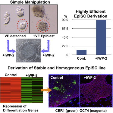
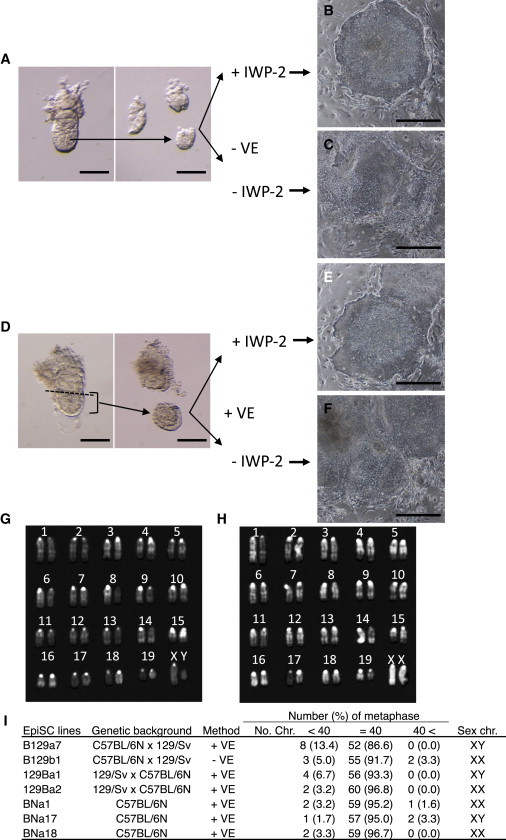
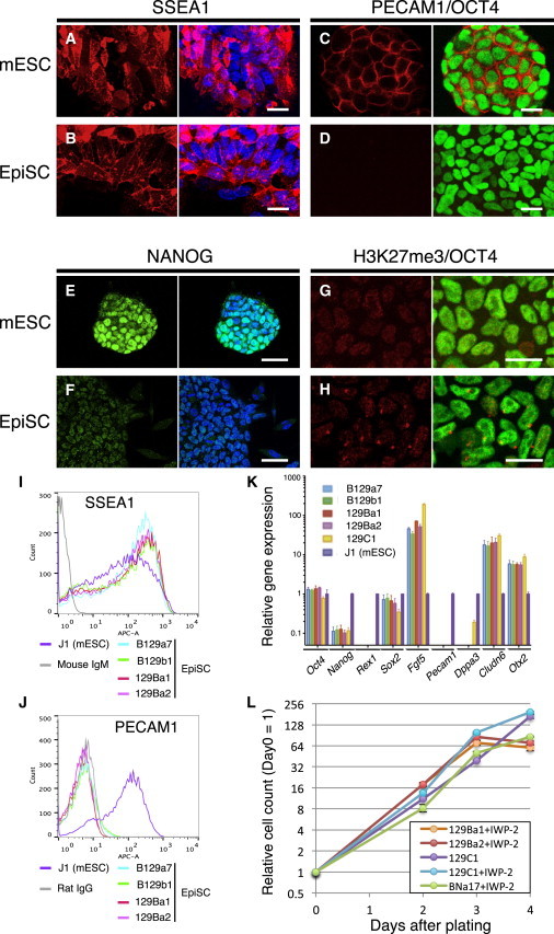
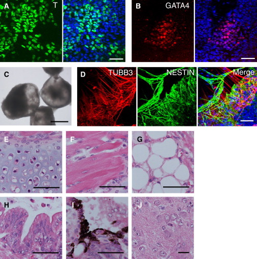
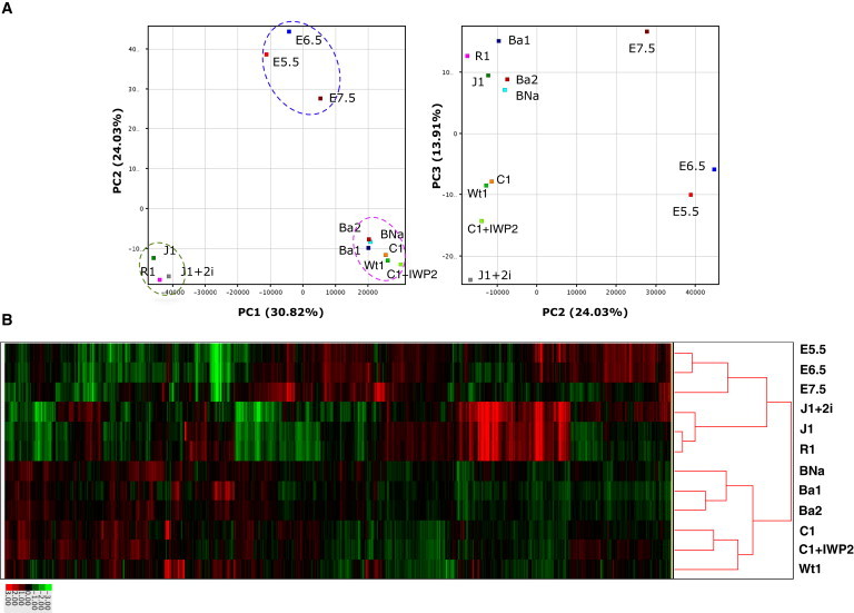
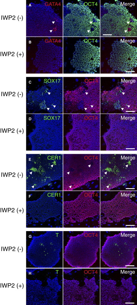
Epiblast stem cells (EpiSCs) are pluripotent stem cells derived from epiblasts of postimplantation mouse embryos, and thus provide a useful model for studying "primed" pluripotent states. Here, we devised a simple and robust technique to derive high-quality EpiSCs using an inhibitor of WNT secretion. Using this method, we readily established EpiSC lines with high efficiency and were able to use whole embryonic portions without having to separate the epiblast from the visceral endoderm (VE). Expression analyses revealed that these EpiSCs maintained a homogeneous, undifferentiated status, yet showed high potential for differentiation both in vitro and in teratomas. Unlike EpiSCs derived by the original protocol, new EpiSC lines required continuous treatment with the Wnt inhibitor, suggesting some intrinsic differences from the existing EpiSCs. The homogeneous properties of this new version of EpiSCs should facilitate studies on the establishment and maintenance of a "primed" pluripotent state, and directed differentiation from the primed state.


Figures

None
Graphical abstract
Figure 1
Figure 1
Establishment of EpiSC Lines by the IWP-2 Method (A) E5.5 epiblasts without VE (−VE) were cultured with or without IWP-2. (B and C) Epiblast outgrowth 5 days after plating of epiblasts without VE and with IWP-2 (B), and without VE and IWP-2 (C). (D) Epiblasts with VE (+VE) of E5.5 embryos were cultured with or without IWP-2. (E and F) Epiblast outgrowth 5 days after plating of epiblasts with VE and IWP-2 (E), and with VE and without IWP-2 (F). (G and H) Representative karyotypes are shown for male (G) and female (H) EpiSC lines. (I) List of the EpiSC lines used in this study, including their genetic background, the derivation method used, chromosome numbers, and compositions of sex chromosomes. Scale bars, 100 μm (A and B) and 0.5 mm (C–F). See also Figure S1.
Figure 2
Figure 2
Cellular Pluripotency Marker Expression Levels in EpiSCs Derived by the IWP-2 Method (A–J) Immunofluorescence images for SSEA1 (A and B), PECAM1 (red) and OCT4 (green, C and D), NANOG (E and F), and histone H3K27 trimethylation (G and H) in mESCs (A, C, E, and G) and EpiSCs (B, D, F, and H). Nuclear staining is shown using TO-PRO3 (blue). Cytograms of EpiSCs and mESCs are shown using anti-SSEA1 (I) and anti-PECAM1 (J) antibodies. (K) Relative expression levels of marker genes in EpiSCs detected by qRT-PCR compared with J1 mESCs. For each gene, technical triplicate assays and two independent experiments were performed. Error bars represent the standard SEM. (L) Growth curves of EpiSCs. At the indicated time points, 2.0 × 104 cells were plated and counted. Averaged data from three independent experiments were plotted. Scale bars, 20 μm (A, B, C, D, G, and H) and 50 μm (E and F).
Figure 3
Figure 3
Differentiation Potential of EpiSCs Isolated by the IWP-2 Method (A and B) Expression levels of Brachyury (T) (A) and GATA4 (B) were detected in B129a4 EpiSCs isolated by the IWP-2 method when they were cultured in medium without IWP-2 for 1 week. (C) Bright-field image of EBs formed from 129Ba2 EpiSCs isolated with IWP-2 treatment. (D) Immunofluorescence images for TUBB3 (red) and NESTIN (green), detecting neural differentiation in 129 Ba2 EBs. (E–J) Hematoxylin and eosin-stained sections of teratomas from 129Ba1 EpiSCs: (E) cartilage, (F) skeletal muscle, (G) adipocytes, (H) gastrointestinal epithelium, (I) melanocytes, and (J) neural tissue. Scale bars, 50 μm (A, B, and D–J) and 0.5 mm (C). See also Figures S2 and S3.
Figure 4
Figure 4
Global Gene-Expression Profiling of EpiSCs, mESCs, and Epiblasts (A) PCA of global gene-expression profiles obtained from the indicated cell types. J1 and R1 are mESCs. J1+2i represents J1 cells cultured in 2i-containing medium. E5.5, E6.5, and E7.5 are epiblast cells isolated from embryos at the corresponding stages. Ba1, Ba2, BNa, Wt1, C1, and C1+IWP2 represent EpiSC lines 129Ba1, 129Ba2, 129BNa, Wt1, 129C1, and 129C1 cultured in the IWP-2-containing medium, respectively. Dotted circles indicate the mESC group (green), EpiSC group (pink), and epiblast group (blue). (B) Hierarchical cluster analysis of expression profiles from EpiSCs, mESCs, and epiblast cells. See also Figure S4 and Table S2.
Figure 5
Figure 5
Effects of IWP-2 on Gene Expression in the 129C1 EpiSC Line Immunofluorescence images for GATA4 (red) and OCT4 (green) (A and B), SOX17 (C and D), T (E and F), and CER1 (G and H) in EpiSCs cultured without IWP-2 (A, C, E, and G) or with IWP-2 (B, D, F, and H). Nuclei were stained with TO-PRO3 (blue). Scale bar, 100 μm. See also Figures S5 and S6.

References

    1. Bao S., Tang F., Li X., Hayashi K., Gillich A., Lao K., Surani M.A. Epigenetic reversion of post-implantation epiblast to pluripotent embryonic stem cells. Nature. 2009;461:1292–1295. - PMC - PubMed
    1. Bernemann C., Greber B., Ko K., Sterneckert J., Han D.W., Araúzo-Bravo M.J., Schöler H.R. Distinct developmental ground states of epiblast stem cell lines determine different pluripotency features. Stem Cells. 2011;29:1496–1503. - PubMed
    1. Biechele S., Cockburn K., Lanner F., Cox B.J., Rossant J. Porcn-dependent Wnt signaling is not required prior to mouse gastrulation. Development. 2013;140:2961–2971. - PubMed
    1. Blauwkamp T.A., Nigam S., Ardehali R., Weissman I.L., Nusse R. Endogenous Wnt signalling in human embryonic stem cells generates an equilibrium of distinct lineage-specified progenitors. Nat. Commun. 2012;3:1070. - PMC - PubMed
    1. Bosma G.C., Custer R.P., Bosma M.J. A severe combined immunodeficiency mutation in the mouse. Nature. 1983;301:527–530. - PubMed

Publication types