

In-line phase-contrast and grating-based phase-contrast synchrotron imaging study of brain micrometastasis of breast cancer
- PMID: 25818989
- PMCID: PMC4377630
- DOI: 10.1038/srep09418
In-line phase-contrast and grating-based phase-contrast synchrotron imaging study of brain micrometastasis of breast cancer
Abstract
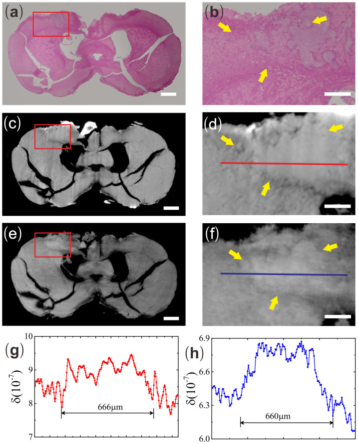
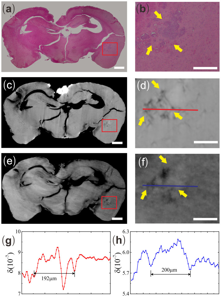
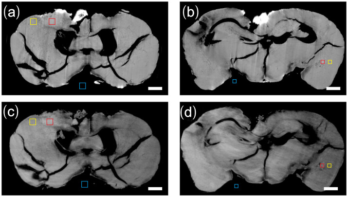
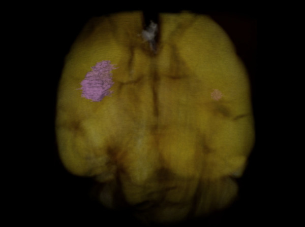
Current bio-medical imaging researches aim to detect brain micrometastasis in early stage for its increasing incidence and high mortality rates. Synchrotron phase-contrast imaging techniques, such as in-line phase-contrast (IPC) and grating-based phase-contrast (GPC) imaging, could provide a high spatial and density imaging study of biological specimens' 3D structures. In this study, we demonstrated the detection efficiencies of these two imaging tools on breast cancer micrometastasis in an ex vivo mouse brain. We found that both IPC and GPC can differentiate abnormal brain structures induced by micrometastasis from the surrounding normal tissues. We also found that GPC was more sensitive in detecting the small metastasis as compared to IPC.
Figures
References
-
- Gupta G. P. & Massagué J. Cancer Metastasis: Building a Framework. Cell. 127, 679–695 (2006). - PubMed
-
- Weigelt B., Peterse J. L. & van't Veer L. J. Breast cancer metastasis: markers and models. Nat Rev Cancer. 5, 591–602 (2005). - PubMed
-
- Melisko M. E. et al. Brain metastases in breast cancer: clinical and pathologic characteristics associated with improvements in survival. J Neurooncol. 88, 359–365 (2008). - PubMed
-
- Shaffrey M. E. et al. Brain metastases. Curr Probl Surg. 41, 665–741 (2004). - PubMed
Publication types
MeSH terms
Substances
LinkOut - more resources
Full Text Sources
Other Literature Sources
Medical
