Review
. 2015 May:479-480:160-6.
doi: 10.1016/j.virol.2015.03.016. Epub 2015 Mar 24.

Necroptosis: The Trojan horse in cell autonomous antiviral host defense

Affiliations
Review

Necroptosis: The Trojan horse in cell autonomous antiviral host defense

Edward S Mocarski et al. Virology. 2015 May.

Abstract

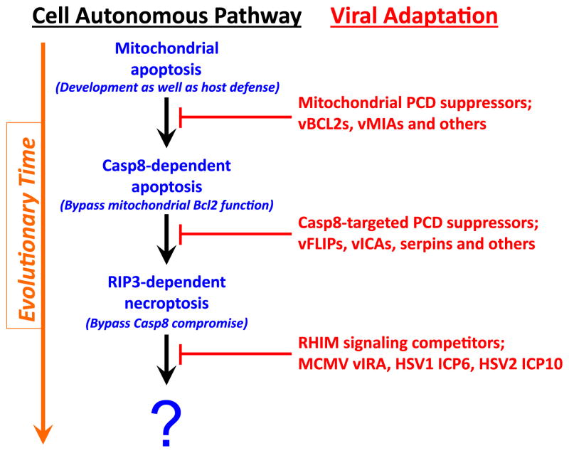
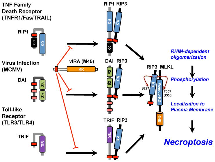
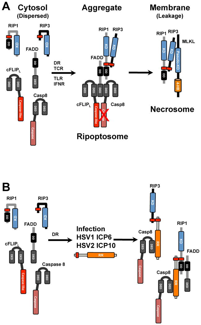
Herpesviruses suppress cell death to assure sustained infection in their natural hosts. Murine cytomegalovirus (MCMV) encodes suppressors of apoptosis as well as M45-encoded viral inhibitor of RIP activation (vIRA) to block RIP homotypic interaction motif (RHIM)-signaling and recruitment of RIP3 (also called RIPK3), to prevent necroptosis. MCMV and human cytomegalovirus encode a viral inhibitor of caspase (Casp)8 activation to block apoptosis, an activity that unleashes necroptosis. Herpes simplex virus (HSV)1 and HSV2 incorporate both RHIM and Casp8 suppression strategies within UL39-encoded ICP6 and ICP10, respectively, which are herpesvirus-conserved homologs of MCMV M45. Both HSV proteins sensitize human cells to necroptosis by blocking Casp8 activity while preventing RHIM-dependent RIP3 activation and death. In mouse cells, HSV1 ICP6 interacts with RIP3 and, surprisingly, drives necroptosis. Thus, herpesviruses have illuminated the contribution of necoptosis to host defense in the natural host as well as its potential to restrict cross-species infections in nonnatural hosts.

Keywords: FADD; Programmed necrosis; Ribonucleotide reductase; Serine/threonine protein kinase; cFLIP.


Figures

Figure 1
Figure 1. Evolutionary relationships in cell autonomous death pathways and virus-encoded countermeasures
Programmed cell death (PCD) pathways include the mitochondrial (or cell-intrinsic) death pathway that eliminates cells during development and homeostatic turnover as well as in host defense [9,10,53,100]. Viral (v)Bcl2 homologs and other virus-encoded mitochondrial suppressors block mitochondrial PCD, establishing conditions that selected for a Casp8-like activity to directly trigger effector caspases, bypassing mitochondrial Bcl2 function. Viruses adaptation included Casp8-targeted cell death suppressors, some specific, like herpesvirus vFLIPs, betaherpesvirus vICA, and some nonspecific, like the poxvirus serpin, CrmA. RIP3-dependent necroptosis via RHIM-dependent interaction with RIP1, TRIF or DAI, enables the host to bypass the impact of virus-encoded Casp8 inhibitors. The examples of MCMV vIRA and HSV1/HSV2 R1 RHIM signaling competitors are the first examples of viral suppressors of necroptosis [2,3,5,20]. Note that HSV1 ICP6 inhibits necroptosis in human cells but promotes necroptosis in mouse cells [98,99]. This depiction is adapted from [9].
Figure 2
Figure 2. Canonical and noncanonical RHIM-dependent activation of RIP3 kinase
Canonical activation of necroptosis via RIP1 recruitment and activation RIP3 downstream of TNF family death receptors (TNFR1, Fas, TRAIL), highlighting the known and predicted conformational changes that accompany this pathway [28]. Noncanonical activation of necroptosis via DAI downstream of MCMV infection or via TRIF downstream of TLR3 and TLR4 activation, driving recruitment and activation of RIP3, with predicted conformational changes. All forms of RHIM-dependent activation of RIP3 are blocked by MCMV-encoded vIRA and converge on the RIP3-dependent recruitment and phosphorylation events leading to plasma membrane leakage and other outcomes characteristic of necroptosis.
Figure 3
Figure 3. Formation and disruption of the ripoptosome and necrosome by HSV1 ICP6 and HSV2 ICP10
A. The ripoptosome forms from cytosolic components. FADD becomes activated downstream of TNF family death receptor (DR) activation, as well as following T cell receptor (TCR), TLR and interferon receptor (IFNR) activation, to recruit Casp8 and cFLIPL heterodimer via homotypic death effector domain (DED) as well as RIP1 via a homotypic death domain (DD) interactions. RIP1 recruits RIP3 via a common RHIM (shown as a red rectangle) and, when Casp8 activity is inhibited, triggers oligomerization that leads to an amyloid-like complex that recruits MLKL into a necrosome that localizes to membranes and directs the final steps in necroptosis leading to membrane leakage. B. Interaction of HSV1 ICP6 and HSV2 ICP10 with ripoptosome components in human cells. Cytosolic ripoptosome components form HSV R1-dependent complexes via RHIM and Casp8 interaction domains discussed in the text.

References

    1. Cho YS, Challa S, Moquin D, Genga R, Ray TD, et al. Phosphorylation-driven assembly of the RIP1-RIP3 complex regulates programmed necrosis and virus-induced inflammation. Cell. 2009;137:1112–1123. - PMC - PubMed
    1. Upton JW, Kaiser WJ, Mocarski ES. DAI (ZBP1/DLM-1) complexes with RIP3 to mediate virus-induced programmed necrosis that is targeted by murine cytomegalovirus vIRA. Cell Host Microbe. 2012;11:290–297. - PMC - PubMed
    1. Upton JW, Kaiser WJ, Mocarski ES. Virus inhibition of RIP3-dependent necrosis. Cell Host Microbe. 2010;7:302–313. - PMC - PubMed
    1. Berger AK, Danthi P. Reovirus activates a caspase-independent cell death pathway. MBio. 2013;4:e00178–00113. - PMC - PubMed
    1. Guo H, Omoto S, Harris PA, Finger JN, Bertin J, et al. Herpes simplex virus suppression of necroptosis in human cells. Cell Host Microbe. 2015;17:243–251. - PMC - PubMed

Publication types