Review
. 2015 Apr:133:19-29.
doi: 10.1016/j.exer.2015.02.011.

Optic nerve head biomechanics in aging and disease

Affiliations
Review

Optic nerve head biomechanics in aging and disease

J Crawford Downs. Exp Eye Res. 2015 Apr.

Abstract

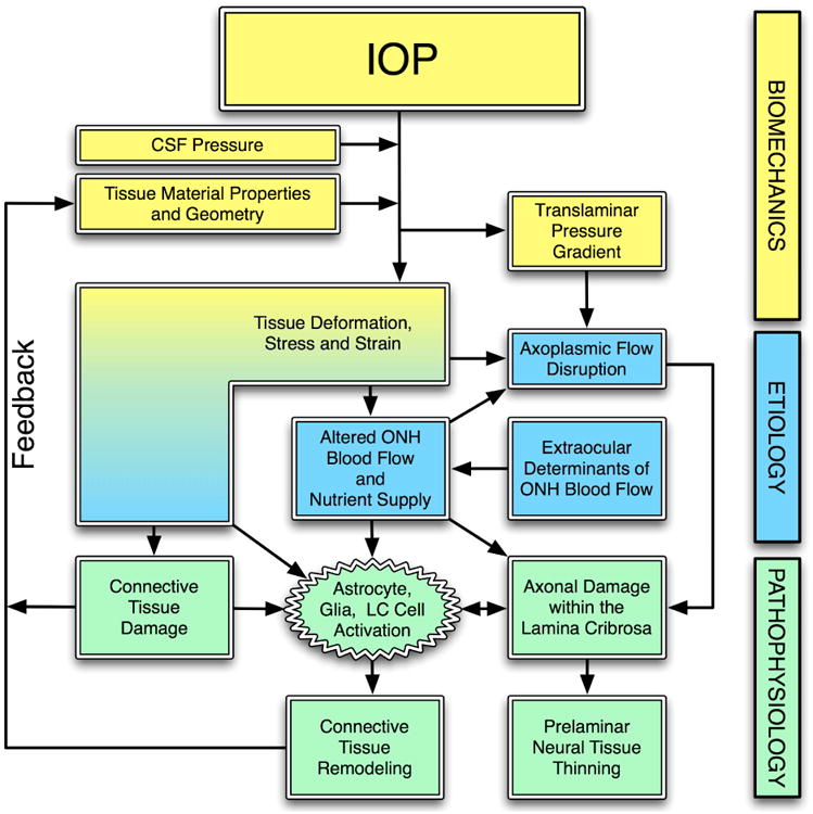
This nontechnical review is focused upon educating the reader on optic nerve head biomechanics in both aging and disease along two main themes: what is known about how mechanical forces and the resulting deformations are distributed in the posterior pole and ONH (biomechanics) and what is known about how the living system responds to those deformations (mechanobiology). We focus on how ONH responds to IOP elevations as a structural system, insofar as the acute mechanical response of the lamina cribrosa is confounded with the responses of the peripapillary sclera, prelaminar neural tissues, and retrolaminar optic nerve. We discuss the biomechanical basis for IOP-driven changes in connective tissues, blood flow, and cellular responses. We use glaucoma as the primary framework to present the important aspects of ONH biomechanics in aging and disease, as ONH biomechanics, aging, and the posterior pole extracellular matrix (ECM) are thought to be centrally involved in glaucoma susceptibility, onset and progression.

Keywords: Biomechanics; Glaucoma; ONH; Optic nerve head; Sclera.


Figures

Figure 1
Figure 1. The optic nerve head (ONH) is a three-dimensional (3D) structure comprised of multiple interactive tissue systems that exist on different scales. This complexity has been a formidable deterrent to characterizing its mechanical environment
(A) While clinicians are familiar with the clinically visible surface of the optic nerve head (referred to as the optic disc), in fact the ONH (B) is a dynamic, 3D structure (seen here in an illustrated sectional view) in which the retinal ganglion cell (RGC) axons in bundles (white) surrounded by glial columns (red), pass through the connective tissue beams of the lamina cribrosa (light blue), isolated following trypsin digestion in an scanning electron micrograph (SEM) of the scleral canal in (C). The blood supply for the connective tissues of the lamina cribrosa (D) derives from the posterior ciliary arteries and the circle of Zinn-Haller (Z-H). (E-F) The relationship of the laminar beams to the axon bundles is shown in schematic form in (E). (F) Individual beams of the lamina cribrosa are lined by astrocytes and LC cells. Together they provide structural and metabolic support for the adjacent axon bundles. Within the lamina, the RGC axons have no direct blood supply. Axonal nutrition requires diffusion of nutrients from the laminar capillaries (solid red), across the endothelial basement membranes, through the extracellular matrix (ECM) of the laminar beam (stippled), across the basement membranes of the astrocytes (thick black), into the astrocytes (yellow), and across their processes (not shown) to the adjacent axons (vertical lines). Chronic age-related changes in the endothelial cell and astrocyte basement membranes, as well as intraocular pressure (IOP)-induced changes in the laminar ECM and astrocyte basement membranes may diminish nutrient diffusion to the axons in the presence of a stable level of laminar capillary volume flow. In advanced glaucoma, the connective tissues of the normal lamina cribrosa (G, sagittal view of the center of the ONH; vitreous above, orbital optic nerve below) change shape and remodel into a cupped and excavated configuration (H). Adapted from (Sigal, Roberts et al. 2010).
Figure 2
Figure 2. IOP-related stress and strain are a constant presence within the ONH at all levels of IOP
IOP and cerebrospinal fluid pressure act mechanically on the tissues of the eye, producing deformations, strain and stress within the tissues. These deformations depend on the eye-specific geometry and material properties of the individual eye. In a biomechanical paradigm, the stress and strain will alter the blood flow (primarily), and the delivery of nutrients (secondarily) through chronic alterations in connective tissue stiffness and diffusion properties. IOP-related stress and strain also induce connective tissue damage directly (laminar beam yield), or indirectly (cell mediated remodeling), which drives a connective tissue remodeling process that alters the tissues' geometry and mechanical response to loading. This feeds back directly onto the mechanical effects of IOP. Adapted from Sigal, Downs, et al. (Sigal, Roberts et al. 2010)
Figure 3
Figure 3. High- and Low-frequency IOP Fluctuation in the Nonhuman primate
Top:Screen capture of approximately 7 seconds of the continuous IOP tracing from an unrestrained awake primate showing baseline mean IOP of ∼8-13 mmHg and IOP fluctuations up to 12 mmHg associated with blinks and saccadic eye movements. IOP fluctuations can be much larger and of longer duration, especially when the animal squints or is agitated or stressed. Bottom: Plot of the 10-minute time-window average of 24 hours of continuous IOP showing low frequency IOP fluctuation from a single nonhuman primate. The color of the plot points and lines indicate how much data were removed from each 10-minute window after post-hoc digital filtering of signal dropout and noise. Green indicates that 100% of the continuous IOP data were used in the 10-minute average IOP plotted in each point, and yellow indicates that 50% were eliminated due to signal dropout or noise. Note the fluctuations in IOP are substantial even when the high-frequency IOP spikes seen in the top plot are averaged out. Adapted from Downs, et al. (Downs, Burgoyne et al. 2011).
Figure 4
Figure 4. Complexities in the Posterior Pole Biomechanical Response
The image on the left shows the strain distribution in a macro-scale model of the connective tissues of the posterior pole of the eye. Note that thickness variations in the sclera give rise to a non-uniform distribution of strain within the scleral shell and that the strains are lower in the sclera than in the more compliant lamina cribrosa. The middle image shows a detail of the strain field within the macro-scale representation of the lamina cribrosa. While this portion of the model has been assigned regional material properties related to the amount and orientation of the laminar beams (based on 3D reconstruction data such as that in Figure 5), the continuum description represents a bulk homogenization of the specific microarchitecture in each element. The right image shows the distribution of mechanical strain at the micro-scale in the laminar beam microarchitecture, demonstrating that strains concentrate focally in individual beams and around individual pores.
Figure 5
Figure 5. Regional differences in laminar microarchitecture in a normal eye, and the predicted relationship between regional laminar density and strain
Characterization of the laminar microarchitecture (Left) utilizes the element boundaries of a continuum finite mesh to partition the lamina cribrosa connective tissue into forty-five sub-regions (Right). The connective tissue volume fraction (CTVF) for each region is expressed as a percentage and mapped to a grayscale value in the background. The arrows indicate the predominant orientation of the laminar beams in each region, with higher values (color-coded) indicating regions in which the beams are more highly oriented. Note that in the peripheral regions of the lamina, the beams are tethered radially into the scleral canal wall. (Bottom) FE model simulations in both eyes of four nonhuman primates show that strains are highest in areas where the laminar density is lowest, and strains are lowest in areas where laminar density is highest (Roberts, Liang et al. 2010). The symbols represent strains in each of the elements, colored by eye and animal, and the lines represent the nonlinear fit to those data.
Figure 6
Figure 6. The Influence of Scleral Mechanics on ONH Mechanics
IOP induces large scleral canal expansions in eyes with compliant sclera (left) that pulls the contained lamina taut despite the direct posterior force of IOP on the laminar surface. Conversely, a stiff sclera allows relatively little canal expansion with IOP elevation (right) and less stretching of the contained lamina, thus allowing the lamina to be displaced posteriorly by the direct action of IOP on its anterior surface. Adapted from (Sigal, Roberts et al. 2010).
Figure 7
Figure 7. A possible link between age-related changes in scleral stiffness and age-related loss of axons in normal eyes?
Maps showing the rate of change in neural retinal rim area measurement with age in normal human eyes (See, Nicolela et al. 2009) and the age-related change in underlying peripapillary scleral structural stiffness (note negative coefficients indicate increased scleral compliance with age and positive coefficients indicate increased stiffness with age) (Fazio, Grytz et al. 2014). While these results are independent and there has not been a causative link established between these phenomena, one might speculate that the age-related changes in scleral stiffness contribute to the pattern of age-related axon loss in normal eyes.

References

    1. Agapova OA, Kaufman PL, Lucarelli MJ, Gabelt BT, Hernandez MR. Differential expression of matrix metalloproteinases in monkey eyes with experimental glaucoma or optic nerve transection. Brain Res. 2003;967(1-2):132–143. - PubMed
    1. Agoumi Y, Sharpe GP, Hutchison DM, Nicolela MT, Artes PH, Chauhan BC. Laminar and prelaminar tissue displacement during intraocular pressure elevation in glaucoma patients and healthy controls. Ophthalmology. 2011;118(1):52–59. - PubMed
    1. Albon J, Karwatowski WS, Avery N, Easty DL, Duance VC. Changes in the collagenous matrix of the aging human lamina cribrosa. Br J Ophthalmol. 1995;79(4):368–375. - PMC - PubMed
    1. Albon J, Purslow PP, Karwatowski WS, Easty DL. Age related compliance of the lamina cribrosa in human eyes. Br J Ophthalmol. 2000;84(3):318–323. - PMC - PubMed
    1. Bailey AJ, Paul RG, Knott L. Mechanisms of maturation and ageing of collagen. Mech Ageing Dev. 1998;106(1-2):1–56. - PubMed

Publication types