. 2015 Apr 20;25(8):1017-30.
doi: 10.1016/j.cub.2015.02.022. Epub 2015 Mar 26.

PLETHORA Genes Control Regeneration by a Two-Step Mechanism

Affiliations

PLETHORA Genes Control Regeneration by a Two-Step Mechanism

Abdul Kareem et al. Curr Biol. .

Abstract

Regeneration, a remarkable example of developmental plasticity displayed by both plants and animals, involves successive developmental events driven in response to environmental cues. Despite decades of study on the ability of the plant tissues to regenerate a complete fertile shoot system after inductive cues, the mechanisms by which cells acquire pluripotency and subsequently regenerate complete organs remain unknown. Here, we show that three PLETHORA (PLT) genes, PLT3, PLT5, and PLT7, regulate de novo shoot regeneration in Arabidopsis by controlling two distinct developmental events. Cumulative loss of function of these three genes causes the intermediate cell mass, callus, to be incompetent to form shoot progenitors, whereas induction of PLT5 or PLT7 can render shoot regeneration hormone-independent. We further show that PLT3, PLT5, and PLT7 establish pluripotency by activating root stem cell regulators PLT1 and PLT2, as reconstitution of either PLT1 or PLT2 in the plt3; plt5-2; plt7 mutant re-established the competence to regenerate shoot progenitor cells but did not lead to the completion of shoot regeneration. PLT3, PLT5, and PLT7 additionally regulate and require the shoot-promoting factor CUP-SHAPED COTYLEDON2 (CUC2) to complete the shoot-formation program. Our findings uncouple the acquisition of competence to regenerate shoot progenitor cells from completion of shoot formation, indicating a two-step mechanism of de novo shoot regeneration that operates in all tested plant tissues irrespective of their origin. Our studies reveal intermediate developmental phases of regeneration and provide a deeper understanding into the mechanistic basis of regeneration.


Figures

Figure 1
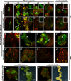
Figure 1. 1. PLT genes are upregulated during shoot regeneration
(A, E’) PLT3::PLT3:vYFP (K, J’) PLT5::PLT5:vYFP and (U, O’) PLT7::PLT7:vYFP expression in untreated LRP and young leaves. (B) Proliferating cells after 3 days of CIM induction are marked with, PLT3-YFP (L) PLT5-YFP and (V) PLT7-YFP. (C–E, M–O, W–Y, F’, G’, K’, L’, P’,Q’) Upregulation of all three PLTs is maintained throughout the callus phase and the expression becomes mostly confined to the sub-epidermal cells of proliferating callus after 7–10 days. (F–H, P–R, Z–B’, H’, M’, R’) Upon SIM treatment the expression of all three PLTs gradually get accumulated in the shoot forming cells in callus. (I) A high expression of PLT3-YFP (S) PLT5-YFP and (C’) PLT7-YFP expression in nascent shoot meristem (arrowhead) after 10 days on SIM. (J, T, D’, I’, N’, S’) All three PLT signals accumulated in leaf primordia (arrow) that emerged from the meristem periphery after 12 days of induction. All images are maximum projections of z-stacks except A, K and U, which are single optical sections. Red signal reflects propidium iodide stain in (A–H, K–R, U–B’), and FM4-64 stain in the remaining. Scale bar: 50 µm in (A,K,U) and 100 µm in the remaining.
Figure 2
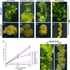
Figure 2. PLT genes are necessary and sufficient for de novo shoot formation
De novo shoot regeneration in wild-type calli derived from (A) leaf, (B) cotyledon, (C) hypocotyl and (D) root explants after 14 days of SIM treatment. (A’–D’) Shoot regeneration is abolished in plt3; plt5-2; plt7 calli derived from leaf, cotyledon, hypocotyl and root explants. (E) Regeneration efficiency in various combinations of plt mutants. Number of shoots were scored per explant and the average length of explant was 3 cm. (F) Shoot regeneration in the callus of wildtype; 35S::PLT5:GR incubated on hormone-free medium supplemented with dexamethasone. (F’) No shoot regeneration in mock-treated callus of wild-type;35S::PLT5:GR. Scale bar: 1mm. Error bar in (E) represents standard error of the mean.
Figure 3
Figure 3. Auxin responses are deregulated in plt3; plt5-2; plt7 mutants after regeneration stimulus
(A,A’) PIN1::PIN1:GFP (green with arrowhead) and pDR5rev::3XVENUS-N7 (yellow) expression in LRP of both wild-type and plt3; plt5-2; plt7 on pre-CIM medium. (B,B’) Upregulation of both DR5-VENUS and PIN1-GFP in proliferating cells of both genotypes after 2 days on CIM.. (C,C’) DR5-VENUS signal is accumulated throughout the proliferating cells after 4 days in both the genotypes while PIN1-GFP (arrowhead) is expressed in the sub-epidermal cells. Inset shows PIN1-GFP expression. (D,E) Downregulation of DR5VENUS and PIN1-GFP in wild-type callus on CIM after 6 and 8 days respectively. Inset shows PIN1-GFP expression. (D’,E’) Downregulation of both DR5VENUS and PIN1-GFP in the mutant after 6 days on CIM and no PIN1-GFP expression is detected after 8 days. (F,F’) Expression of DR5VENUS and PIN1-GFP (arrowhead) in leaf-derived calli of both wild-type and mutant after 6 days on CIM. Inset shows DR5VENUS signal (G) After 6 days on SIM, sporadic distribution of DR5VENUS signal and no PIN1-GFP expression in wild-type. (H) Upregulation of PIN1-GFP in developing-shoot meristem (arrowhead) in wild-type after 10 days on SIM. DR5VENUS is expressed within the initiating leaf primordia and in the peripheral callus. (I) Both PIN1-GFP and DR5VENUS signal accumulated within leaf primordia (arrow) after 12 days. (J) Upregulation of PIN1-GFP and DR5VENUS fluorescence in leaf primordia (arrow) occurred in the callus derived from leaf explants on SIM. (G’) Weak expression of DR5-VENUS in plt3; plt5-2; plt7 after 6 days on SIM. (H’-J’) No PIN1-GFP expression and a weak and ubiquitous expression of DR5VENUS without any localized signal accumulation in the mutant calli derived from both root and leaf explants after 10–12 days on SIM. (K) PIN1-GFP is localized the tip of leaf primordia (arrow) in wild-type;pDR5::PIN1:GFP after 10 days on SIM. (K’) No shoot meristem formation in plt3; plt5-2; plt7;pDR5::PIN1:GFP although a weak PIN1-GFP expression is observable in most parts of the callus. (L) Shoot regeneration in wild-type;pDR5::PIN1:GFP after 12 days on SIM. (L’) No shoot regeneration in plt3; plt5-2; plt7;pDR5::PIN1:GFP on SIM. (S) The panels (A and A’) are confocal single optical section images, (L and L’) are bright field images and the remaining are confocal images with projections of multiple optical sections. Red colour is the propidium iodide stain in (A–F’) and the FM4-64 stain in (G,G’-J’),. Red colour in (H,I) is autofluorescence. Scale bar= 50µm in (A–J’, K,K’) and 1 mm in (L,L’)
Figure 4
Figure 4. WUS and CLV3 expression domains are not properly established in plt3; plt5-2; plt7 mutants after regeneration stimulus
(A) 2 days after transfer to SIM, WUS::erCFP was expressed in the innermost layers of proliferating cells in wild-type. (B,C) WUS expression was distributed in a large portion of the wild-type callus after 4–6 days and (D,E) it became progressively restricted to the centre of developing meristems. Inset in (D) shows WUS expression in the meristem centre. (F) After 12 days, WUS-CFP marked the centre of shoot meristems in wild-type. (A’) In plt3; plt5-2; plt7 callus, WUS was weakly expressed after 2 days of SIM induction and (B’-D’) became scattered within the callus. (E’,F’) Scattered distribution continued after 10–12 days of induction, without any confined accumulation. (G) CLV3::erCFP expression initially arose in wild-type callus after 2 days of SIM induction. (H–K) After 4–10 days of induction, CLV3::erCFP signal expanded to encompass a large part of wild-type callus. (L) CLV3::erCFP was upregulated only in the meristem centre after 12 days of SIM induction in wild-type. (G’) Weak expression of CLV3::erCFP in plt3; plt5-2; plt7 callus after 2 days of induction. (H’-K’) Sporadic CLV3::erCFP expression in plt3; plt5-2; plt7 callus after 4–10 days of SIM induction. (L’) CLV3::erCFP signal remained sporadic without any localized upregulation after 12 days of induction. (M,N) Ectopic overexpression of WUS (G10-90::WUS:3AT) in wild-type induced de novo shoots from both callus and LRP upon incubation on hormone free medium supplemented with estradiol. (M’,N’) Overexpression of WUS in plt3; plt5-2; plt7 mutant tissue did not induce de novo shoots on callus or LRP. (O) Forced induction of ESR2 (G10-90::ESR2:3AT) on minimal medium with estradiol induced de novo shoots on wild-type callus while (O’) mutant callus failed to regenerate shoots. Scale bar: 50µm in (A–L’); 1mm in (M–O’).
Figure 5
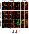
Figure 5. Root stem cell maintenance regulators are not detectably expressed in plt3; plt5-2; plt7 LRP and callus
(A–F, G–L, M–R) pSCR::H2B:YFP, pPLT2::PLT2:YFP, and pWER::H2B:YFP expression in wild-type explants and (A’–F’, G’–L’, M’–R’) plt3; plt5-2; plt7 explants. The order of the columns from the left is: untreated primary root tip, untreated lateral root primordium, calli derived from root cultured on CIM for 5 days, 11days, and calli derived from leaf cultured on CIM for 3 days, 6 days. While in wild-type explants, all three reporters are expressed in both untreated primary root tip, LRP, and CIM-induced calli derived from root and leaf (A–F, G–L, M–R), in the plt3; plt5-2; plt7 mutant explants, pSCR::H2B:YFP and pPLT2::PLT2:YFP are expressed neither in LRP (B’, H’ asterisks) nor calli derived from those tissues (C’–F’, I’–L’). (N’) The LRP of plt3; plt5-2; plt7 displays slight pWER::H2B:YFP expression. (P’, Q’, R’) In calli of plt3; plt5-2; plt7, partial or weak expression is observed. (O’) Some callus does not express the reporter at all. (S) Upregulation of PLT1 and PLT2 transcripts upon the induction of PLT5 measured by quantitative RT-PCR. Expression levels were normalized to ACTIN2. Error bar represents standard error of the mean from three independent biological replicates. Scale bar in (A–R’) = 50µm.
Figure 6
Figure 6. Root stem cell maintenance regulators establish early competence for shoot regeneration
(A) Shoot regeneration in wild-type callus (Wild-type;PLT7::PLT1:YFP) derived from root explant after 12 days on SIM. (B,E) Competent calli turned green in plt3; plt5-2; plt7;PLT7::PLT1:vYFP root and leaf explants and in (C) plt3; plt5-2; plt7;PLT3::PLT2:GR root explants after 12 days on SIM. (D,F) Callus derived from root and leaf explants of plt3; plt5-2; plt7 remained yellowish on SIM. (G) Expression of PIN1-GFP in Wild-type; PIN1::PIN1:GFP after 7 days on CIM. (H) plt3; plt5-2; plt7;PLT7::PLT1:vYFP, PIN1::PIN1:GFP callus regained cellular morphology typical of wild-type and expressed PIN1-GFP after 7 days on CIM (I) Disorganized callus cells without PIN1-GFP expression in plt3; plt5-2; plt7;PIN1::PIN1:GFP on CIM. (J) Shoot progenitor cells labeled with PIN1-GFP in wild-type callus and (K) in plt3; plt5-2; plt7;PLT7::PLT1:vYFP, PIN1::PIN1:GFP after 7 days on SIM (L) No PIN1-GFP expression or shoot progenitor cell formation in plt3; plt5-2; plt7;PIN1::PIN1:GFP on SIM. Scale bar: 1mm in (A–F) and 50µm in (G–L). Red color in (G–I) is propidium iodide. No stain was used for cell boundaries in (J–L).
Figure 7
Figure 7. PLT3, PLT5 and PLT7 control de novo shoot regeneration by a two-step mechanism
(A) Bar graph showing CUC1 and CUC2 expression levels in wild-type and plt3; plt5-2; plt7 mutant calli after 10 days of induction on SIM, measured by quantitative RT-PCR. (B) pCUC2::3X-VENUS was upregulated in wild-type callus on SIM and (C) it was downregulated in plt3; plt5-2; plt7 callus. (D) CUC1 and CUC2 transcripts levels after 4 hrs of PLT5 induction by DEX and (E) DEX with cycloheximide treatment, measured by quantitative RT-PCR. Expression levels were normalized to ACTIN2. (F) Bar graph showing percentage of shoots formed in wild-type;35S::PLT5:GR and cuc1-5,cuc2- 3;35S::PLT5:GR after 4 weeks of culture on hormone free medium supplemented with DEX. (G) De novo shoot regeneration is completely abolished in plt3; plt5-2; plt7 upon SIM induction. (H) Upon reconstitution of PLT1 expression in plt3; plt5-2; plt7, callus cells regain pluripotency and shoot progenitors are regenerated. Arrowheads represent green foci. (I) De novo shoot formation is not achieved in plt3; plt5-2; plt7; 35S::CUC2 upon SIM treatment (J) Ectopic overexpression of CUC2 in plt3; plt5-2; plt7;PLT7::PLT1-YFP leads to complete shoot regeneration on SIM. (K) Schematic representation of a two-step mechanism of shoot regeneration. First, PLT3, PLT5 and PLT7 control the expression of root stem cell maintenance regulators enabling regenerative competence and second, they regulate shoot promoting factors leading to the initiation of shoot regeneration. (K–i) Explants derived from aerial or root tissues (K-ii) PLT3, PLT5 and PLT7 determine pluripotency by regulating the root stem cell maintenance regulators PLT1 and PLT2 (K-iii) Pluripotent callus can regenerate shoot progenitor cells on SIM. Root stem cell maintenance regulators are downregulated on SIM. (K-iv) Shoot progenitor cells further require shoot-promoting factors (CUC2) regulated by PLT3, PLT5 and PLT7 to complete the process of shoot regeneration. Error bars in (A,D,E) represent standard error of the mean from three independent biological replicates. Scale bar in (B,C) = 50µm and in (G–I) = 1mm

References

    1. Birnbaum KD, Sanchez Alvarado A. Slicing across kingdoms: regeneration in plants and animals. Cell. 2008;132:697–710. - PMC - PubMed
    1. Pulianmackal AJ, Kareem AV, Durgaprasad K, Trivedi ZB, Prasad K. Competence and regulatory interactions during regeneration in plants. Frontiers in plant science. 2014;5:142. - PMC - PubMed
    1. Gordon SP, Heisler MG, Reddy GV, Ohno C, Das P, Meyerowitz EM. Pattern formation during de novo assembly of the Arabidopsis shoot meristem. Development (Cambridge, England) 2007;134:3539–3548. - PubMed
    1. Atta R, Laurens L, Boucheron-Dubuisson E, Guivarc'h A, Carnero E, Giraudat-Pautot V, Rech P, Chriqui D. Pluripotency of Arabidopsis xylem pericycle underlies shoot regeneration from root and hypocotyl explants grown in vitro. The Plant journal : for cell and molecular biology. 2009;57:626–644. - PubMed
    1. Skoog F, Miller CO. Chemical regulation of growth and organ formation in plant tissues cultured in vitro. Symposia of the Society for Experimental Biology. 1957;11:118–130. - PubMed

Publication types

MeSH terms