. 2015 Mar 27;4(4):420-34.
doi: 10.1242/bio.201411072.

Bridging the gap between postembryonic cell lineages and identified embryonic neuroblasts in the ventral nerve cord of Drosophila melanogaster

Affiliations

Bridging the gap between postembryonic cell lineages and identified embryonic neuroblasts in the ventral nerve cord of Drosophila melanogaster

Oliver Birkholz et al. Biol Open. .

Abstract

The clarification of complete cell lineages, which are produced by specific stem cells, is fundamental for understanding mechanisms, controlling the generation of cell diversity and patterning in an emerging tissue. In the developing Central Nervous System (CNS) of Drosophila, neural stem cells (neuroblasts) exhibit two periods of proliferation: During embryogenesis they produce primary lineages, which form the larval CNS. After a phase of mitotic quiescence, a subpopulation of them resumes proliferation in the larva to give rise to secondary lineages that build up the CNS of the adult fly. Within the ventral nerve cord (VNC) detailed descriptions exist for both primary and secondary lineages. However, while primary lineages have been linked to identified neuroblasts, the assignment of secondary lineages has so far been hampered by technical limitations. Therefore, primary and secondary neural lineages co-existed as isolated model systems. Here we provide the missing link between the two systems for all lineages in the thoracic and abdominal neuromeres. Using the Flybow technique, embryonic neuroblasts were identified by their characteristic and unique lineages in the living embryo and their further development was traced into the late larval stage. This comprehensive analysis provides the first complete view of which embryonic neuroblasts are postembryonically reactivated along the anterior/posterior-axis of the VNC, and reveals the relationship between projection patterns of primary and secondary sublineages.

Keywords: CNS development; Cell lineage; Drosophila; Flybow; Neuroblast; Segmental patterning.


Conflict of interest statement

Competing interests: The authors declare no competing or financial interests.

Figures

Fig. 1.
Fig. 1.. Generation and Processing of Clonal Data.
(A–B′″) Lineage tracing using DiI labelling (Bossing et al., 1996b; Schmidt et al., 1997; A–A′″) as compared to applying Flybow 1.1B (Shimosako et al., 2014; B–B′″). (A) Schematic transverse sections showing right ventral half of a stage 6 and a stage 16 embryo; ventral is down, mesoderm in mint, mesectoderm and neuroectoderm are in beige. Classical labelling of single neuroectodermal progenitor cells (stage 6, capillary with red dye from right) allows targeted application of the dye (e.g. dorsal versus ventral cells or thoracic versus abdominal segments) and results in complete lineages at the end of embryogenesis (stage 16). (B) Schematic transverse sections showing right ventral half of a stage 10 and a stage 16 embryo (elav>Flybow 1.1B). Independent recombination events in (1) a NB, (2) a ganglion mother cell and (3) a postmitotic neuron, caused by a heatshock driven flipase are symbolised with arrowheads (stage 10). In stage 16 these events result in multicellular (1), two cell (2) or single cell clones (3), respectively, labelled randomly by mCherry-, mCitrine- or mTurquoise-expression. Maximum clone size and -density can be controlled by the timepoint and strength of the heatshock, but clone locations are arbitrary. Corresponding clonal types labelled by both methods can be unambiguously identified in vivo based on typical morphological characteristics: compare (A′–A′″) and (B′–B′″) respectively (dorsal views of early stage 17 VNCs). CBG, cell body glia; CG, channel glia; Ic, Ica, Icp, Ii, TB are specific interneurons (see Bossing et al., 1996b; Schmidt et al., 1997). (C–F) Demonstration of the Flybow 1.1B approach used to record, process and present the larval clone data. (C,D) Dorsal views on thoracic segments of a late L3 larva, representing the unprocessed stack that was used for Fig. 3B. (C′) resembles the default state (GFP-expression) in all neuronal cells without recombination event, while (C) extracts the three channels resulting from recombination. The yellow channel reveals only single cell clones, red shows a MNB- and a NB4-2 clone, cyan a NB4-1 clone. (C″) Nrt-antibody staining, visualising commissural fibres, that served as landmarks to identify secondary lineages (see text and Fig. 2 for details). (D) If a clone was chosen for presentation, the respective channel (here: the red one) was transformed into grey. Then every single layer was inspected and secondary cells and their projections were coloured red. Primary cells and all remaining projections clearly connected to the clone were coloured green (for discrimination between primary and secondary cells see text). (E–E″″) Stacks of several images from ventral (E) to dorsal (E″″) positions, documenting the procedure that results in (F), i.e. the complete stack and the same image as shown in Fig. 3B. White vertical bars indicate the midline.
Fig. 2.
Fig. 2.. Pattern of Neurotactin-positive fibre bundles.
Horizontal view of projections (several sections) through the ventral (A), intermediate (B) and dorsal (C) neuropil of the thorax and through the ventral neuropil of the abdomen (D) of a late L3 larva of the indicated genotype, stained against Nrt (magenta). GFP was recorded only weakly to not interfere with the Nrt-staining. The characteristic Nrt-positive bundles (indicated on the left) serve as landmarks to identify postembryonic cell lineages (see Truman et al., 2004) and corresponding segments. For abbreviations see text. Asterisks mark the leg neuropils, which are additionally highlighted by stippled lines. White vertical bars indicate the midline.
Fig. 3.
Fig. 3.. Characteristics of MNB-, NB1-1- and NB1-2-lineage.
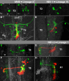
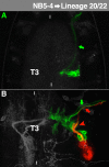
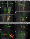
(A,A′,C,E) Horizontal view (several sections) of the thoracic VNC of living embryos. (B,D,F,G,H) Horizontal view (maximum projection) of the thoracic (B,D,F,G) or anterior abdominal VNC (H) of fixed late third larval instars. In this figure and Figs 4–11, all embryonic images are marked by green, all larval ones by white capital letters. The primary sublineages are illustrated in green and the secondary sublineages in red, which does not reflect their original fluorophore expression (see Materials and Methods). Clone characteristics are highlighted by symbols (green: embryonic origin; red: larval origin; filled arrowhead: ipsilateral projections; hollow arrowhead: contralateral projections, filled arrow: motorprojections, hollow arrow: glial cells) and introduced in the text. Segments are indicated. The background fluorescence of other clones is in grey and allows orientation. White bars indicate the midline. Please see text for morphological details.
Fig. 4.
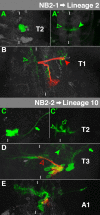
Fig. 4.. Characteristics of NB2-1- and NB2-2-lineage.
For details, see the text and the legend of Fig. 3.
Fig. 5.
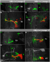
Fig. 5.. Characteristics of NB2-4-, NB2-5- and NB3-1-lineage.
For details, see the text and the legend of Fig. 3.
Fig. 6.
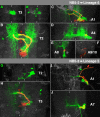
Fig. 6.. Characteristics of NB3-2-, NB3-3-, NB3-4-, NB3-5- and NB4-1-lineage.
For details, see the text and the legend of Fig. 3.
Fig. 7.
Fig. 7.. Characteristics of NB4-2-, NB4-3- and NB4-4-lineage.
For details, see the text and the legend of Fig. 3.
Fig. 8.
Fig. 8.. Characteristics of NB5-2- and NB5-3-lineage.
For details, see the text and the legend of Fig. 3.
Fig. 9.
Fig. 9.. Characteristics of NB5-4-lineage.
For details, see the text and the legend of Fig. 3.
Fig. 10.
Fig. 10.. Characteristics of NB6-1- and NB6-2-lineage.
For details, see the text and the legend of Fig. 3.
Fig. 11.
Fig. 11.. Characteristics of NB6-4-, NB7-1- and NB7-4-lineage.
For details, see the text and the legend of Fig. 3.
Fig. 12.
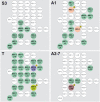
Fig. 12.. Segment-specific pattern and identities of embryonic neuroblasts, which are reactivated in the larva.
Right hemineuromeres of the indicated segments are schematically shown. In each NB its embryonic name (upper row) according to Broadus et al. (Broadus et al., 1995) and Doe (Doe, 1992) and its postembryonic cell lineage (lower row) according to Brown and Truman (Brown and Truman, 2009), Kuert et al. (Kuert et al., 2014) and Truman et al. (Truman et al., 2004) are indicated, except for A2-A7, where the lower row indicates the name of the postembryonic progenitor according to Truman and Bate (Truman and Bate, 1988). NBs which are reactivated in the larva, are highlighted in green; those which are mitotically inactive are white; other colours mark NBs with segment-specific characteristics: (1) Lineage 18 was not identified in T1. (2) Lineage 11 was not identified in T3. (3) NB2-2 and NB5-3 only form a few secondary neurons in A1 and reveal PCD within their lineage. (4) NB6-2 is only reactivated in A2, but not posterior to it.

References

    1. Almeida M. S., Bray S. J. (2005). Regulation of post-embryonic neuroblasts by Drosophila Grainyhead. Mech. Dev. 122, 1282–1293 10.1016/j.mod.2005.08.004 - DOI - PubMed
    1. Baek M., Mann R. S. (2009). Lineage and birth date specify motor neuron targeting and dendritic architecture in adult Drosophila. J. Neurosci. 29, 6904–6916 10.1523/JNEUROSCI.1585-09.2009 - DOI - PMC - PubMed
    1. Baumgardt M., Karlsson D., Terriente J., Díaz-Benjumea F. J., Thor S. (2009). Neuronal subtype specification within a lineage by opposing temporal feed-forward loops. Cell 139, 969–982 10.1016/j.cell.2009.10.032 - DOI - PubMed
    1. Bello B. C., Hirth F., Gould A. P. (2003). A pulse of the Drosophila Hox protein Abdominal-A schedules the end of neural proliferation via neuroblast apoptosis. Neuron 37, 209–219 10.1016/S0896-6273(02)01181-9 - DOI - PubMed
    1. Bello B., Holbro N., Reichert H. (2007). Polycomb group genes are required for neural stem cell survival in postembryonic neurogenesis of Drosophila. Development 134, 1091–1099 10.1242/dev.02793 - DOI - PubMed

LinkOut - more resources