. 2015 Feb 19;59(1):2459.
doi: 10.4081/ejh.2015.2459.

Elastofibroma dorsi: a histochemical and immunohistochemical study

Affiliations

Elastofibroma dorsi: a histochemical and immunohistochemical study

A Di Vito et al. Eur J Histochem. .

Abstract

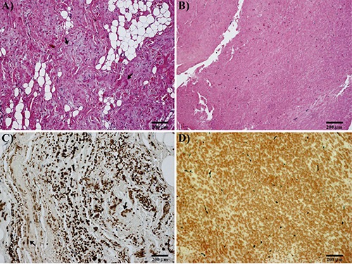
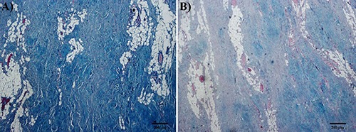
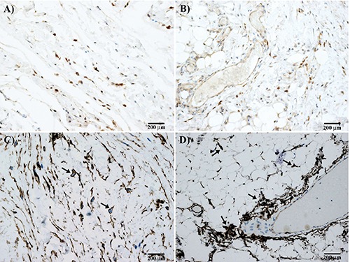
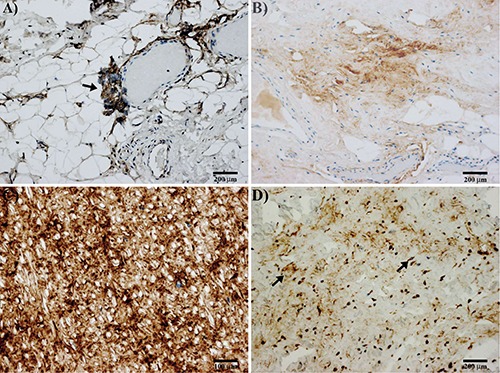
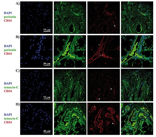
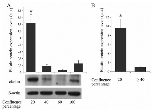
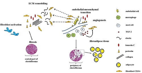
Elastofibroma dorsi (ED) is considered a member of a heterogeneous group of benign fibrous (fibroblastic or myofibroblastic) soft-tissue tumors, frequently localized in the periscapular region in middle aged or older individuals. However, the pathogenesis of ED is still unclear and many authors believe that ED results from a reactive hyperproliferation of fibroblastic tissue, while others suggest that it may be a consequence of a mechanical friction. In our study, we examined 11 cases of ED using histochemical and immunohistochemical methods, in order to extend the knowledge about extracellular matrix composition and histopathogenesis of ED. From the results it appeared that stroma and interspersed spindle cells of ED were positive for both periostin and tenascin-C. Mast cells tryptase-positive were also abundant throughout the lesion. The perivascular distribution of periostin and tenascin-C, associated with the CD34 positivity, suggest that endothelial-mesenchymal transition events can account for neovascularization and production of fibroelastic tissue characteristic of elastofibroma. Our data obtained in endothelial cells cultures demonstrated that elastin production is higher when the status of confluence of the cells is low. So, we can assume that such a phenomenon is a characteristic of mesenchymal/endothelial cells CD34 positive, in which elastin production results to be inversely proportional to the vascular differentiation of cellular elements. In the light of these considerations, we think that a cancerous nature of ED is unlikely. Overall, our study report, for the first time, a detailed description of extracellular matrix composition in ED, suggesting that a mechanical strain-dependent reactivation of periostin and tenascin-C expression, as well as of elastin deposition, could be responsible for development of ED.


Conflict of interest statement

Conflicts of interest: the authors declare no conflicts of interest.

Figures

Figure 1.
Figure 1.
A) Microscopic findings of elastofibroma; elastic connective is intermixed with fibrous tissue (H&E; arrows indicating elastic fibers). B) Ligamentum flavum: note the regular and monotonous structure (H&E). C) Elastofibroma; immunohistochemical stain for elastic fibers: note the irregular arrangement of coarse elastic fibers (arrows). D) Ligamentum flavum; immunohistochemical stain for elastic fibers: note the uniform arrangement of elastic fibers.
Figure 2.
Figure 2.
Results of special stains of elastofibroma. A) Alcian Blue pH 2.5. B) Alcian Blue pH 2.5 after treatment with testicular hyaluronidase. Considerable amount of hyaluronic acid is observed in the lesion.
Figure 3.
Figure 3.
Immunohistochemical results of elastofibroma. Dark brown stained mast cells were positive for tryptase in central fibrous (A) and peripheral vascularized (B) area of elastofibroma. Many fibroblasts are also positive for CD34 in central (C; arrows) and peripheral (D) area of elastofibroma. Note in (D) the perivascular but also scattered distribution of cells in the fibroadipose tissue (arrows).
Figure 4.
Figure 4.
Immunohistochemical results of elastofibroma. A) Immunostaining for periostin in peripheral vascularized part of elastofibroma; note the staining of an angiogenetic perivascular tuft of cells, sign of an endothelial-mesenchymal transition (arrow). B) Immunostaining for periostin in the central area of elastofibroma; note the lower positivity for periostin in the stroma. C) Strong staining for periostin is observed in ligamentum flavum. D) Immunostaining for tenascin-C in elastofibroma; tenascin-C positivity in the stroma and cells scattered in central part of elastofibroma (arrows); perivascular positivity for tenascin-C is also observed in the peripheral part of the lesion (not shown).
Figure 5.
Figure 5.
Colocalization of CD34 and mesenchymal markers periostin or tenascin-C. Double immunofluorescence staining with the antibodies against periostin (green) and CD34 (red) in central (A) and peripheral (B) area of the elastofibroma dorsi. Double immunofluorescence staining with the antibodies against tenascin-C (green) and CD34 (red) in central (C) and peripheral (D) area of the elastofibroma dorsi; arrows indicate the positivity for CD34 in interspersed cells, while asterisks indicate the perivascular positivity for CD34.
Figure 6.
Figure 6.
A,B) Levels of elastin in HUVEC cell cultures at different confluence densities; a.u., arbitrary unit; *P<0.05.
Figure 7.
Figure 7.
Elastofibroma pathogenesis. Chronic mechanical strain or genetic predisposition account for excessive fibroblast activation and accumulation of ECM components. Specific positional cues also induce endothelial-mesenchymal transition events. Extravasating macrophages and mast cells develop in the tissue and contribute to fibroblasts activation. In a later stages, the persistent presence of these cells and profibrotic stimuli such as TGF-β, account for progressive development of fibrosis and fat accumulation. A considerable amount of abnormal elastic fibers is observed in the central part of the lesion, where CD34-positive fibroblasts grown at low density.

References

    1. El Hammoumi M, Qtaibi A, Arsalane A, El Oueriachi F, Kabiri el H. Elastofibroma dorsi: clinicopathological analysis of 76 cases. Korean J Thorac Cardiovasc Surg 2014;47:111-6. - PMC - PubMed
    1. Hisaoka M, Nishio JN. Elastofibroma, p. 53-4. Fletcher DM, Bridge JA, Hogendoorn CW, Mertens F.(eds.), WHO classification of tumors of soft tissue and bone. International Agency for research on cancer, Lyon: 2013.
    1. Karakurt O, Kaplan T, Gunal N, Gulbahar G, Kocer B, Han S, et al. Elastofibroma dorsi management and outcomes: review of 16 cases. Interact Cardiovasc Thorac Surg 2014;18:197-201. - PMC - PubMed
    1. Naylor MF, Nascimento AG, Sherrick AD, McLeod RA. Elastofibroma dorsi: radiologic findings in 12 patients. AJR Am J Roentgenol 1996;167:683-7. - PubMed
    1. Schick S, Zembsch A, Gahleitner A, Wanderbaldinger P, Amann G, Breitenseher M, et al. Atypical appearance of elastofibroma dorsi on MRI: case reports and review of the literature. J Comput Assist Tomogr 2000;24:288-92. - PubMed

MeSH terms

LinkOut - more resources