

The fuss over lipo"fuss"cin: not all autofluorescence is the same
- PMID: 25820568
- PMCID: PMC4378222
- DOI: 10.4081/ejh.2015.2512
The fuss over lipo"fuss"cin: not all autofluorescence is the same
Abstract
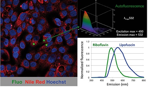
Since the first description of cellular autofluorescence over a century ago, we have now come to appreciate that autofluorescence should not be discarded as a biological artifact but embraced as a biological phenomenon with potentially important cellular relevance. Indeed, cellular and tissue autofluorescence has been attributed to a spectrum of unrelated molecules such as porphyrins, vitamins (vitamin A, riboflavin, thiamine), structural proteins, lipofuscin and ceroid pigments. We have recently shown that freshly isolated epithelial cancer stem cells (CSCs) bear autofluorescent vesicles in the cytoplasm. Our studies definitively prove that riboflavin and not lipofuscin is the source of autofluorescence in CSCs as the inhibition of ATP and not autophagy eliminates CSC autofluorescence, that the ATP-dependent transporter ABCG2, for which riboflavin is a substrate, is overexpressed in autofluorescent CSCs and co-localizes with the membrane of intracellular autofluorescent vesicles, the ABCG2-specific inhibitor Fumitremorgin C reversibly eliminates CSC autofluorescence, riboflavin is a substrate for ABCG2, and only the addition of riboflavin to vitamin-deprived CSC cultures is capable of restoring autofluorescence. Thus, the sum of these data unequivocally supports the conclusion that the source of CSC autofluorescence is the vitamin riboflavin.
Conflict of interest statement
Conflict of interest: the authors declare that no conflict of interests exists for the presented study.
Figures
Comment on
-
Lipofuscin, lipofuscin-like pigments and autofluorescence.Eur J Histochem. 2015 Feb 6;59(1):2485. doi: 10.4081/ejh.2015.2485. Eur J Histochem. 2015. PMID: 25820564 Free PMC article. Review.
References
-
- Miranda-Lorenzo I, Dorado J, Lonardo E, Alcala S, Serrano AG, Clausell-Tormos J, et al. Intracellular autofluorescence: a biomarker for epithelial cancer stem cells. Nat Methods 2014;11:1161-9. - PubMed
-
- Wolman M. Lipid pigments (chromolipids): their origin, nature, and significance. Pathobiol Annu 1980;10:253-67. - PubMed
-
- Brunk UT, Jones CB, Sohal RS. A novel hypothesis of lipofuscinogenesis and cellular aging based on interactions between oxidative stress and autophagocytosis. Mutat Res 1992;275):395-403. - PubMed
Publication types
MeSH terms
Substances
LinkOut - more resources
Full Text Sources
Other Literature Sources
