. 2015 May 1;194(9):4362-70.
doi: 10.4049/jimmunol.1402384. Epub 2015 Mar 27.

Regulatory T cells require TCR signaling for their suppressive function

Affiliations

Regulatory T cells require TCR signaling for their suppressive function

Amanda M Schmidt et al. J Immunol. .

Abstract

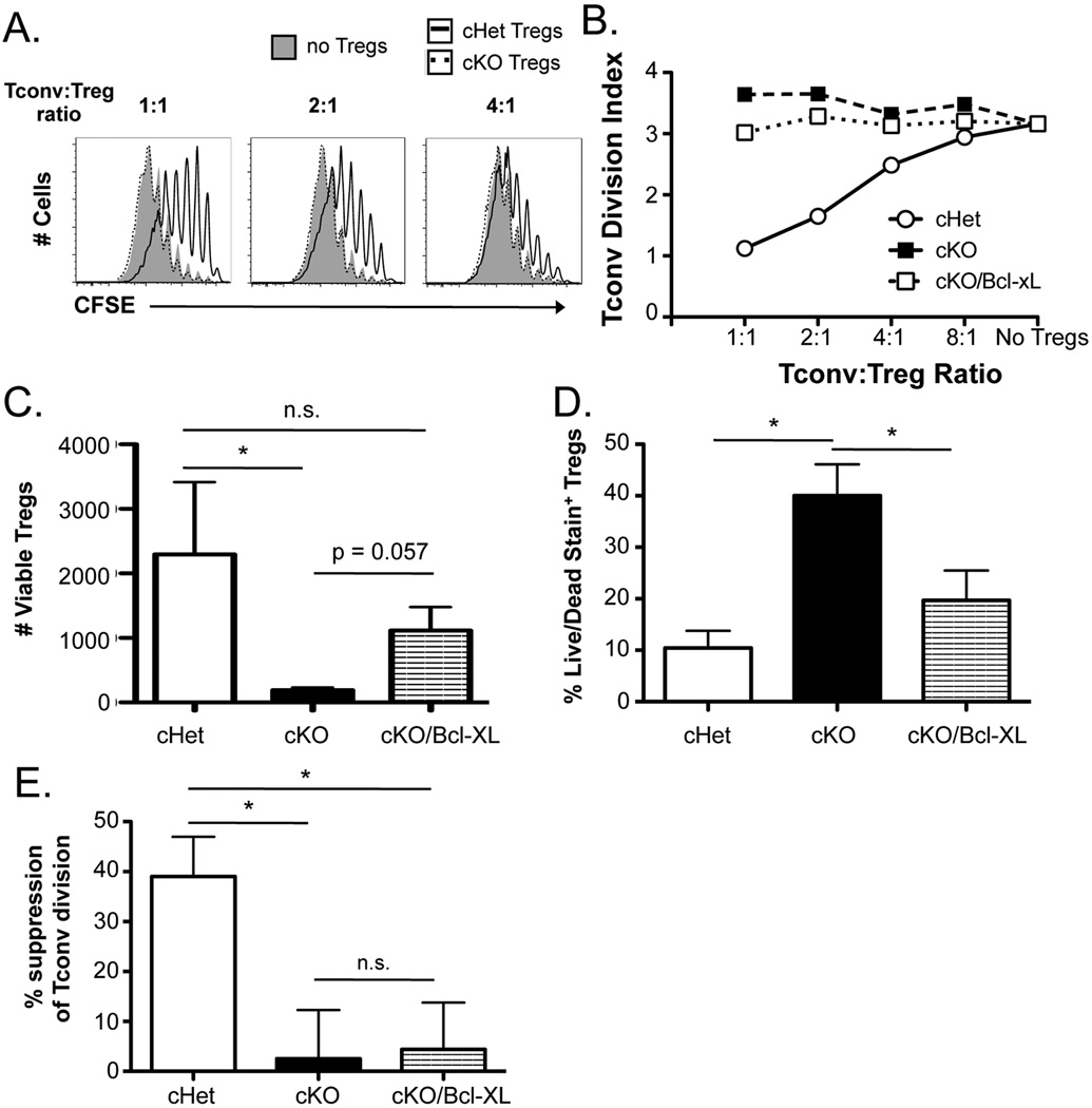
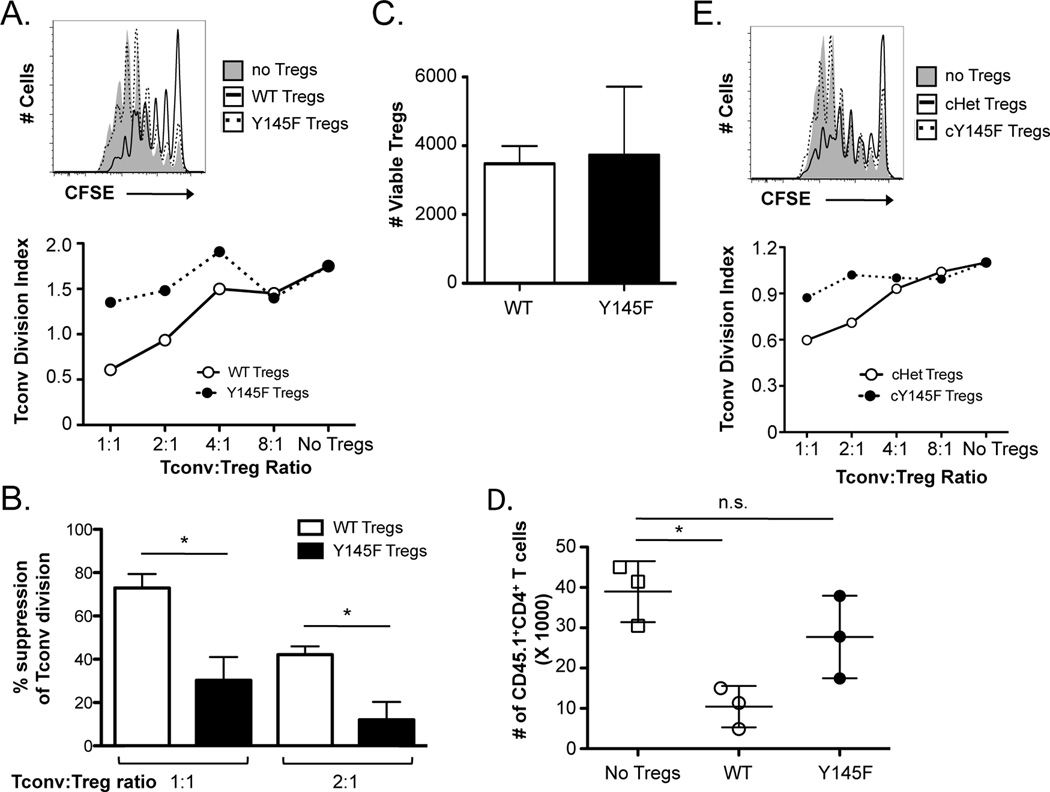
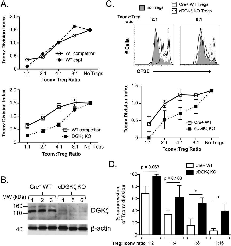
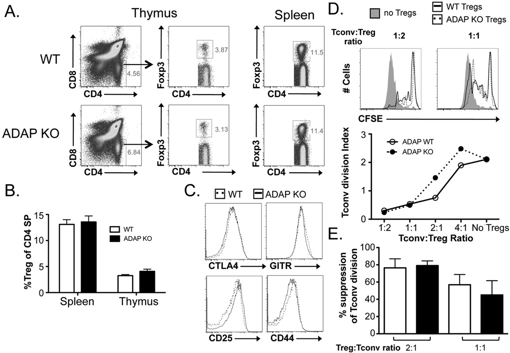
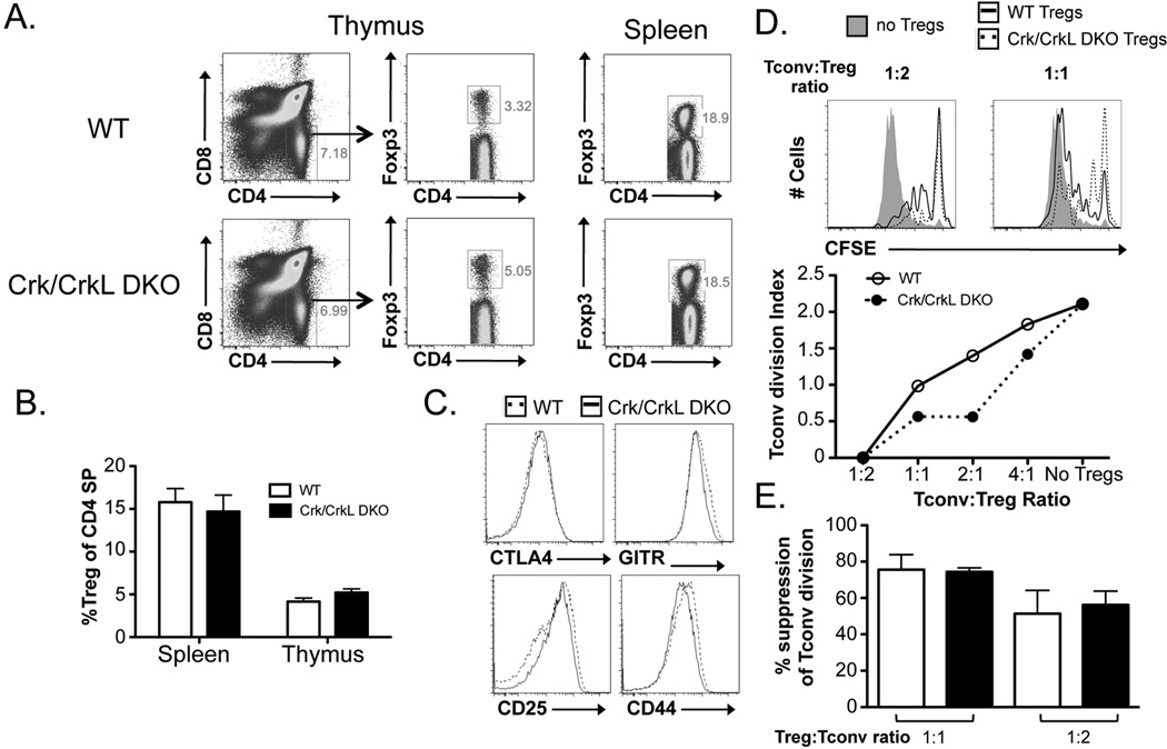
Regulatory T cells (Tregs) are a subset of CD4(+) T cells that maintain immune tolerance in part by their ability to inhibit the proliferation of conventional CD4(+) T cells (Tconvs). The role of the TCR and the downstream signaling pathways required for this suppressive function of Tregs are not fully understood. To yield insight into how TCR-mediated signals influence Treg suppressive function, we assessed the ability of Tregs with altered TCR-mediated signaling capacity to inhibit Tconv proliferation. Mature Tregs deficient in Src homology 2 domain containing leukocyte protein of 76 kDa (SLP-76), an adaptor protein that nucleates the proximal signaling complex downstream of the TCR, were unable to inhibit Tconv proliferation, suggesting that TCR signaling is required for Treg suppressive function. Moreover, Tregs with defective phospholipase C γ (PLCγ) activation due to a Y145F mutation of SLP-76 were also defective in their suppressive function. Conversely, enhancement of diacylglycerol-mediated signaling downstream of PLCγ by genetic ablation of a negative regulator of diacylglycerol kinase ζ increased the suppressive ability of Tregs. Because SLP-76 is also important for integrin activation and signaling, we tested the role of integrin activation in Treg-mediated suppression. Tregs lacking the adaptor proteins adhesion and degranulation promoting adapter protein or CT10 regulator of kinase/CT10 regulator of kinase-like, which are required for TCR-mediated integrin activation, inhibited Tconv proliferation to a similar extent as wild-type Tregs. Together, these data suggest that TCR-mediated PLCγ activation, but not integrin activation, is required for Tregs to inhibit Tconv proliferation.


Conflict of interest statement

Conflict of Interest

The authors declare no conflicts of interest.

Figures

Figure 1
Figure 1. SLP-76-deficient Tregs display no suppressive ability in vitro
(A) CFSE-labeled splenic CD4+Foxp3 cells (Tconvs) were co-cultured with YFP+CD4+CD25+ Tregs from Tamoxifen-treated SLP-76 cHet, SLP-76 cKO, or SLP-76 cKO/Bcl-xL mice for four days in the presence of irradiated T cell-depleted splenocytes and anti-CD3. Representative CFSE plots from cultures lacking Tregs or containing a 1:1, 2:1, or 4:1 Tconv:Treg ratio are shown, and (B) the division index of Tconvs at various Tconv:Treg ratios is shown. (C) The absolute number of live and (D) the fraction of live/dead viability stain-positive SLP-76 cHet, SLP-76 cKO, and SLP-76 cKO/Bcl-xL Tregs from 4 independent experiments were determined using a viability dye in the 4-day co-cultures. (E) The division index of Tconvs at a 2:1 Tconv:Treg ratio was normalized to the baseline Tconv division (no Tregs) for each experiment (n = 4 experiments) and represented as mean % suppression ± SEM. n.s. = not significant, * indicates statistical significance of p < 0.05 by Mann Whitney test or unpaired two-tailed Student t-test.
Figure 2
Figure 2. SLP-76 Y145F Tregs demonstrate greatly diminished in vitro and in vivo suppressive function
(A) CD4+CD25+ Tregs were sorted from SLP-76 Y145F mice and co-cultured with CFSE-labeled CD4+Foxp3 cells (Tconvs) at various ratios for four days in the presence of irradiated T cell-depleted splenocytes and anti-CD3. A representative CFSE plot from cultures lacking Tregs or at a 1:1 Treg:Tconv ratio is shown (top). A representative plot of the division index of Tconvs at various Treg:Tconv ratios is shown (bottom). (B) The percent suppression at a 1:1 and 1:2 Treg:Tconv ratio were compiled from 3 independent experiments and represented as mean ± SEM. (C) The absolute number of live WT and SLP-76 Y145F Tregs from 3 independent experiments was determined using a viability dye in the 4-day co-cultures. (D) TCRβ/δ DKO mice were adoptively transferred with Tconvs with or without Tregs from B6 or SLP-76 Y145F mice. The absolute number of Tconvs recovered is shown. One representative of two independent experiments is shown. (E) YFP+CD4+CD25+ Tregs from Tamoxifen-treated SLP-76 cHet or SLP-76 cY145F mice were co-cultured with CFSE-labeled Tconvs for four days in the presence of irradiated T cell-depleted splenocytes and anti-CD3. A CFSE plot from cultures lacking Tregs or at a 1:1 Treg:Tconv ratio (top) and a plot of the division index of Tconvs at various Treg:Tconv ratios are shown (bottom). One representative of 2 independent experiments is shown. * indicates statistical significance of p < 0.05 by unpaired two-tailed Student t-test.
Figure 3
Figure 3. DGKζ KO Tregs exhibit enhanced suppressive capacity
(A) CD4+CD25+ Tregs were sorted from WT/WT or WT/DGKζ KO mixed BM chimeras. WT competitor Tregs (CD45.1+/CD45.2+) or experimental Tregs (CD45.2+; WT or DGKζ KO) were cultured with CFSE-labeled Tconvs for four days in the presence of irradiated T cell-depleted splenocytes and anti-CD3. The division index of Tconvs at various Treg:Tconv ratios is shown for cells sorted from WT/WT (top) and WT/DGKζ KO mixed BM chimeras (bottom). The data are represented as mean ± SEM of n = 3 mixed BM chimeras. One representative of 2 independent experiments is shown. (B) FACS-sorted YFP+CD4+ T cells from Tamoxifen-treated T2-Cre+ WT (lanes 1–3) or cDGKζ KO mice (lanes 4–6) were FACS-sorted, lysed, and analyzed for expression for DGKζ and β-actin by Western blot analysis. (C) YFP+CD4+CD25+ Tregs from Tamoxifen-treated T2-Cre+ WT and cDGKζ KO mice were co-cultured with CFSE-labeled Tconvs for four days in the presence of irradiated T cell-depleted splenocytes and anti-CD3. Representative CFSE plots from four-day cultures lacking Tregs or at a 2:1 or 8:1 Tconv:Treg ratio of either Cre+ WT or cDGKζ KO Tregs (top) and a plot of the division index of Tconvs at various Tconv:Treg ratios are shown (bottom). The data are represented as mean ± SEM of n = 3 separate mice (bottom). One representative of 2 independent experiments is shown. (D) The division index of Tconvs at various Tconv:Treg ratios were normalized to the baseline Tconv division (no Tregs) for each experiment (n = 2 experiments) and represented as mean % suppression ± SEM (n = 4 mice total). * indicates statistical significance of p<0.05 by unpaired two-tailed Student t-test.
Figure 4
Figure 4. ADAP KO mice harbor Tregs with normal development and suppressive function
(A) Representative flow cytometric plots and (B) the fraction of Tregs (out of CD4+ T cells) from the thymus and spleen of WT and ADAP KO Tregs are shown. (C) The expression of CTLA-4, CD25, GITR, and CD44 on Tregs from WT and ADAP KO Tregs is shown. (D) CFSE-labeled splenic CD4+Foxp3 Tconvs were cultured with either WT or ADAP KO Tregs for four days in the presence of irradiated T cell-depleted splenocytes and anti-CD3. Representative CFSE plots from four-day cultures lacking Tregs or containing a 1:2 or 1:1 Tconv to Treg ratio of either WT or ADAP KO Tregs are shown (top). A plot of the division index of Tconvs at various Treg:Tconv ratios are shown (middle). (E) The division index of Tconvs at Tconv:Treg ratios of 1:2 and 1:1 were normalized to the baseline Tconv division (no Tregs) for each experiment and represented as mean % suppression ± SEM (n = 3 mice total).
Figure 5
Figure 5. Crk/CrkL DKO Tregs do not show decreased suppressive function compared to WT Tregs
(A) Representative flow cytometric plots and (B) the fraction of Tregs (out of CD4+ T cells) from the thymus and spleen of WT and Crk/CrkL DKO Tregs are shown. (C) The expression of CTLA-4, CD25, GITR, and CD44 on Tregs from WT and Crk/CrkL DKO Tregs is shown. (D) CFSE-labeled splenic CD4+Foxp3 Tconvs were cultured with either WT or Crk/CrkL DKO Tregs for four days in the presence of irradiated T cell-depleted splenocytes and anti-CD3. Representative CFSE plots from four-day cultures lacking Tregs or containing a 1:2 or 1:1 Tconv to Treg ratio of either WT or Crk/CrkL DKO Tregs are shown (top). A plot of the division index of Tconvs at various Treg:Tconv ratios are shown (middle). (E) The division index of Tconvs at Tconv:Treg ratios of 1:2 and 1:1 were normalized to the baseline Tconv division (no Tregs) for each experiment and represented as mean % suppression ± SEM (n = 4 mice total).

References

    1. Brunkow ME, Jeffery EW, Hjerrild KA, Paeper B, Clark LB, Yasayko SA, Wilkinson JE, Galas D, Ziegler SF, Ramsdell F. Disruption of a new forkhead/winged-helix protein, scurfin, results in the fatal lymphoproliferative disorder of the scurfy mouse. Nat Genet. 2001;27:68–73. - PubMed
    1. Kim JM, Rasmussen JP, Rudensky AY. Regulatory T cells prevent catastrophic autoimmunity throughout the lifespan of mice. Nature immunology. 2007;8:191–197. - PubMed
    1. Lahl K, Loddenkemper C, Drouin C, Freyer J, Arnason J, Eberl G, Hamann A, Wagner H, Huehn J, Sparwasser T. Selective depletion of Foxp3+ regulatory T cells induces a scurfy-like disease. The Journal of experimental medicine. 2007;204:57–63. - PMC - PubMed
    1. Sakaguchi S, Sakaguchi N, Asano M, Itoh M, Toda M. Immunologic self-tolerance maintained by activated T cells expressing IL-2 receptor alpha-chains (CD25). Breakdown of a single mechanism of self-tolerance causes various autoimmune diseases. Journal of immunology. 1995;155:1151–1164. - PubMed
    1. Belkaid Y, Rouse BT. Natural regulatory T cells in infectious disease. Nature immunology. 2005;6:353–360. - PubMed

Publication types

MeSH terms