Review
. 2015 Mar 13:6:117.
doi: 10.3389/fimmu.2015.00117. eCollection 2015.

Lipid and Protein Co-Regulation of PI3K Effectors Akt and Itk in Lymphocytes

Affiliations
Review

Lipid and Protein Co-Regulation of PI3K Effectors Akt and Itk in Lymphocytes

Xinxin Wang et al. Front Immunol. .

Abstract

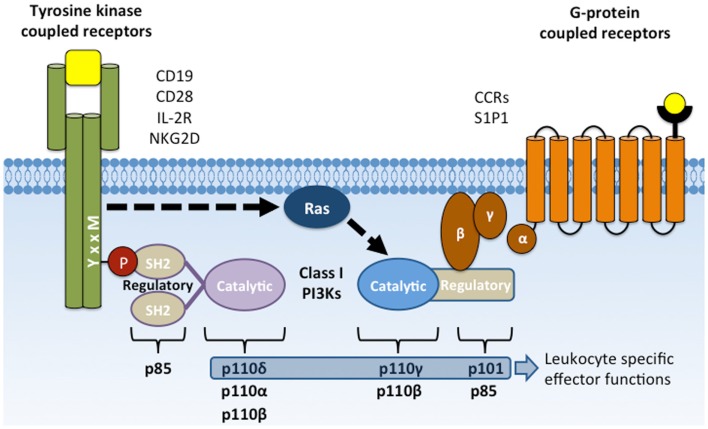
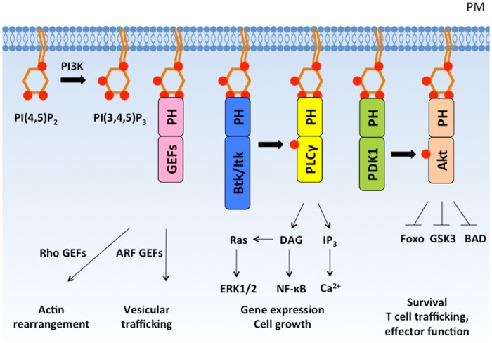
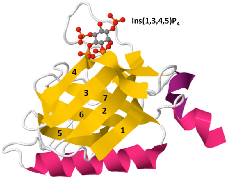
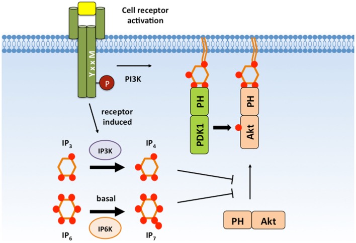
The phosphoinositide 3-kinase (PI 3-kinase, PI3K) pathway transduces signals critical for lymphocyte function. PI3K generates the phospholipid PIP3 at the plasma membrane to recruit proteins that contain pleckstrin homology (PH) domains - a conserved domain found in hundreds of mammalian proteins. PH domain-PIP3 interactions allow for rapid signal propagation and confer a spatial component to these signals. The kinases Akt and Itk are key PI3K effectors that bind PIP3 via their PH domains and mediate vital processes - such as survival, activation, and differentiation - in lymphocytes. Here, we review the roles and regulation of PI3K signaling in lymphocytes with a specific emphasis on Akt and Itk. We also discuss these and other PH domain-containing proteins as they relate more broadly to immune cell function. Finally, we highlight the emerging view of PH domains as multifunctional protein domains that often bind both lipid and protein substrates to exert their effects.

Keywords: Akt signaling; Itk signaling; PI3K; lymphocyte activation; pleckstrin homology domain.


Figures

Figure 1
Figure 1
Activation of class I PI3Ks by YxxM signaling subunits and GPCRs. Membrane receptors that activate PI3K include CD19, CD28, and NKG2D co-receptors, cytokine receptors (e.g., IL-2R), G-protein-coupled receptors (chemokine receptors), and Fcγ receptor I and III. Class IA PI3Ks are recruited to the plasma membrane through SH2 domain interactions with phosphorylated YxxM motifs. Class IB PI3Ks are recruited and activated by direct interaction with the Gβγ subunit following GPCR activation. Activated PI3K phosphorylates the membrane lipid PI(4,5)P2 to form PI(3,4,5)P3.
Figure 2
Figure 2
PI(3,4,5)P3 recruits PH domain-containing proteins to the plasma membrane and regulates diverse cellular responses. PI3K phosphorylates PI(4,5)P2 to form PI(3,4,5)P3, which recruits PH domain-containing signaling proteins to the plasma membrane. PH domain-containing proteins are activated at the plasma membrane and mediate important cellular responses such as cytoskeleton rearrangement, cell growth, proliferation, and survival. PM, plasma membrane; GEF, guanine nucleotide exchange factor.
Figure 3
Figure 3
Crystal structure of Btk PH domain in complex with Ins(1,3,4,5)P4 (PDB ID: 2Z0P). The PH domain is comprised of a β-barrel formed by seven β-strands (yellow, 1–7) capped by an α-helix (pink). The hyper-variable loops of the β-barrel form the binding surface for lipid ligands such as Ins(1,3,4,5)P4 [top, shown by ball-and-stick model: red (oxygen), orange (phosphorus), and gray (carbon)].
Figure 4
Figure 4
IP4 and IP7 negatively regulate Akt signaling. IP4 and IP7 are cytosolic PIP3 analogs that are able to associate with the Akt PH domain with high affinity and can compete with membrane PIP3. IP4 and IP7 binding has been proposed to dissociate Akt from the plasma membrane to prevent Akt activation and substrate accessibility. IP4, Ins(1,3,4,5)P4; IP7, 5-PP-(1,2,3,4,6)IP5; PIP3, PI(3,4,5)P3.
Figure 5
Figure 5
PH domain interactions stabilize Vav1 auto-inhibition in basal state. In the basal state, Vav1 adopts an auto-inhibitory conformation in which the substrate-docking site within the DH domain is blocked by interactions with a helix region from the Ac domain. The interactions between CH, PH, and Ac domains greatly strengthen the auto-inhibitory conformation (left). During T cell activation, phosphorylation of the Ac domain by Lck releases the substrate-docking site and allows GTPase binding (right).
Figure 6
Figure 6
CaM binds the Itk PH domain in a positive feedback loop that potentiates Itk activity, intracellular Ca2+ release, and IL-17A production. Binding of Itk to PIP3 promotes Itk activation and the subsequent phosphorylation and activation of PLC γ1. PLCγ1 cleaves PIP2 to produce DAG and IP3, which binds IP3 receptors on the ER. The IP3 receptor is a ligand-gated Ca2+ channel, and its activation increases Ca2+ levels in the cytosol. Increased cytosolic Ca2+ activates CaM, which has at least two effects on T cell activation: (1) Ca2+/CaM binds to Itk’s PH domain, enhancing its interaction with PIP3 and Itk activity. (2) Ca2+/CaM binds to and activates calcineurin, a phophatase that dephosphorylates NFAT, allowing NFAT translocation to the nucleus where it drives the transcription of IL-17A. Thus, CaM binding to Itk’s PH domain completes a positive feedback loop that potentiates the downstream effects of Itk. PM, plasma membrane; ER, endoplasmic reticulum; Itk, IL-2-inducible tyrosine/T cell kinase; PLCγ1, phospholipase C gamma 1; CaM, calmodulin; NFAT, nuclear factor of activated T cells; IP3R, IP3 receptor.

Similar articles

Cited by

References

    1. Gadina M, Sudarshan C, Visconti R, Zhou YJ, Gu H, Neel BG, et al. The docking molecule gab2 is induced by lymphocyte activation and is involved in signaling by interleukin-2 and interleukin-15 but not other common gamma chain-using cytokines. J Biol Chem (2000) 275:26959–66.10.1074/jbc.M004021200 - DOI - PubMed
    1. Ward SG, Cantrell DA. Phosphoinositide 3-kinases in T lymphocyte activation. Curr Opin Immunol (2001) 13:332–8.10.1016/S0952-7915(00)00223-5 - DOI - PubMed
    1. Koyasu S. The role of PI3K in immune cells. Nat Immunol (2003) 4:313–9.10.1038/ni0403-313 - DOI - PubMed
    1. Guthridge MA, Lopez AF. Phosphotyrosine/phosphoserine binary switches: a new paradigm for the regulation of PI3K signalling and growth factor pleiotropy? Biochem Soc Trans (2007) 35:250–2.10.1042/BST0350250 - DOI - PubMed
    1. Swainson L, Kinet S, Mongellaz C, Sourisseau M, Henriques T, Taylor N. IL-7-induced proliferation of recent thymic emigrants requires activation of the PI3K pathway. Blood (2007) 109:1034–42.10.1182/blood-2006-06-027912 - DOI - PubMed