

Negative energy balance induced by paradoxical sleep deprivation causes multicompartmental changes in adipose tissue and skeletal muscle
- PMID: 25821467
- PMCID: PMC4364052
- DOI: 10.1155/2015/908159
Negative energy balance induced by paradoxical sleep deprivation causes multicompartmental changes in adipose tissue and skeletal muscle
Abstract
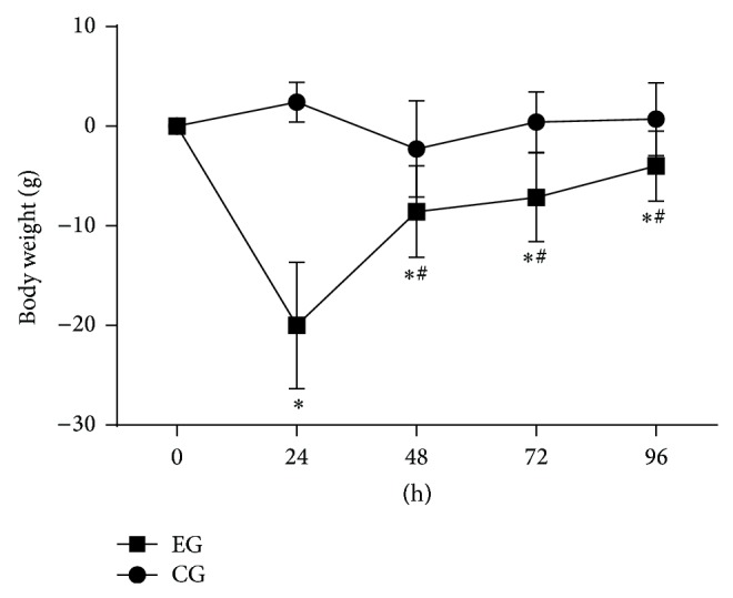
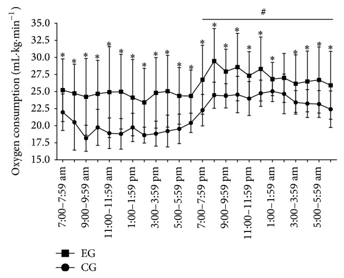
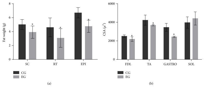
Objective. Describe multicompartmental changes in the fat and various muscle fiber types, as well as the hormonal profile and metabolic rate induced by SD in rats. Methods. Twenty adult male Wistar rats were equally distributed into two groups: experimental group (EG) and control group (CG). The EG was submitted to SD for 96 h. Blood levels of corticosterone (CORT), total testosterone (TESTO), insulin like growth factor-1 (IGF-1), and thyroid hormones (T3 and T4) were used to assess the catabolic environment. Muscle trophism was measured using a cross-sectional area of various muscles (glycolytic, mixed, and oxidative), and lipolysis was inferred by the weight of fat depots from various locations, such as subcutaneous, retroperitoneal, and epididymal. The metabolic rate was measured using oxygen consumption ([Formula: see text]O2) measurement. Results. SD increased CORT levels and decreased TESTO, IGF-1, and T4. All fat depots were reduced in weight after SD. Glycolytic and mixed muscles showed atrophy, whereas atrophy was not observed in oxidative muscle. Conclusion. Our data suggest that glycolytic muscle fibers are more sensitive to atrophy than oxidative fibers during SD and that fat depots are reduced regardless of their location.
Figures
Similar articles
-
Histopathological changes and oxidative damage in type I and type II muscle fibers in rats undergoing paradoxical sleep deprivation.Cell Signal. 2021 May;81:109939. doi: 10.1016/j.cellsig.2021.109939. Epub 2021 Jan 30. Cell Signal. 2021. PMID: 33529759
-
Resistance training minimizes catabolic effects induced by sleep deprivation in rats.Appl Physiol Nutr Metab. 2015 Nov;40(11):1143-50. doi: 10.1139/apnm-2015-0061. Epub 2015 Jul 21. Appl Physiol Nutr Metab. 2015. PMID: 26513007
-
Leucine supplementation is anti-atrophic during paradoxical sleep deprivation in rats.Amino Acids. 2016 Apr;48(4):949-957. doi: 10.1007/s00726-015-2142-7. Epub 2015 Dec 8. Amino Acids. 2016. PMID: 26645537
-
Paradoxical sleep deprivation induces muscle atrophy.Muscle Nerve. 2012 Mar;45(3):431-3. doi: 10.1002/mus.22322. Muscle Nerve. 2012. PMID: 22334180
-
Glucocorticoid-induced skeletal muscle atrophy.Int J Biochem Cell Biol. 2013 Oct;45(10):2163-72. doi: 10.1016/j.biocel.2013.05.036. Epub 2013 Jun 24. Int J Biochem Cell Biol. 2013. PMID: 23806868 Review.
Cited by
-
The effect of sleep restriction, with or without high-intensity interval exercise, on myofibrillar protein synthesis in healthy young men.J Physiol. 2020 Apr;598(8):1523-1536. doi: 10.1113/JP278828. Epub 2020 Mar 11. J Physiol. 2020. PMID: 32078168 Free PMC article.
-
Sleep deprivation regulates availability of PrPC and Aβ peptides which can impair interaction between PrPC and laminin and neuronal plasticity.J Neurochem. 2020 May;153(3):377-389. doi: 10.1111/jnc.14960. Epub 2020 Feb 5. J Neurochem. 2020. PMID: 31950499 Free PMC article.
-
Cytokines, Masticatory Muscle Inflammation, and Pain: an Update.J Mol Neurosci. 2020 May;70(5):790-795. doi: 10.1007/s12031-020-01491-1. Epub 2020 Feb 1. J Mol Neurosci. 2020. PMID: 32008162 Review.
-
Sleep and memory: The impact of sleep deprivation on transcription, translational control, and protein synthesis in the brain.J Neurochem. 2023 Jul;166(1):24-46. doi: 10.1111/jnc.15787. Epub 2023 Mar 25. J Neurochem. 2023. PMID: 36802068 Free PMC article. Review.
-
Acute sleep loss results in tissue-specific alterations in genome-wide DNA methylation state and metabolic fuel utilization in humans.Sci Adv. 2018 Aug 22;4(8):eaar8590. doi: 10.1126/sciadv.aar8590. eCollection 2018 Aug. Sci Adv. 2018. PMID: 30140739 Free PMC article. Clinical Trial.
References
-
- Hipólide D. C., Suchecki D., de Carvalho Pinto A. P., Faria E. C., Tufik S., Luz J. Paradoxical sleep deprivation and sleep recovery: effects on the hypothalamic-pituitary-adrenal axis activity, energy balance and body composition of rats. Journal of Neuroendocrinology. 2006;18(4):231–238. doi: 10.1111/j.1365-2826.2006.01412.x. - DOI - PubMed
-
- Martins P. J. F., Marques M. S., Tufik S., D'Almeida V. Orexin activation precedes increased NPY expression, hyperphagia, and metabolic changes in response to sleep deprivation. The American Journal of Physiology—Endocrinology and Metabolism. 2010;298(3):E726–E734. doi: 10.1152/ajpendo.00660.2009. - DOI - PubMed
-
- Mathur P. P., Chattopadhyay S. Effect of sleep deprivation on the physiological status of rat testis. Andrologia. 1991;23(1):49–51. - PubMed
LinkOut - more resources
Full Text Sources
Other Literature Sources
Miscellaneous
