. 2015:2015:458123.
doi: 10.1155/2015/458123. Epub 2015 Mar 4.

Age-related alterations in the expression of genes and synaptic plasticity associated with nitric oxide signaling in the mouse dorsal striatum

Affiliations

Age-related alterations in the expression of genes and synaptic plasticity associated with nitric oxide signaling in the mouse dorsal striatum

Aisa N Chepkova et al. Neural Plast. 2015.

Abstract

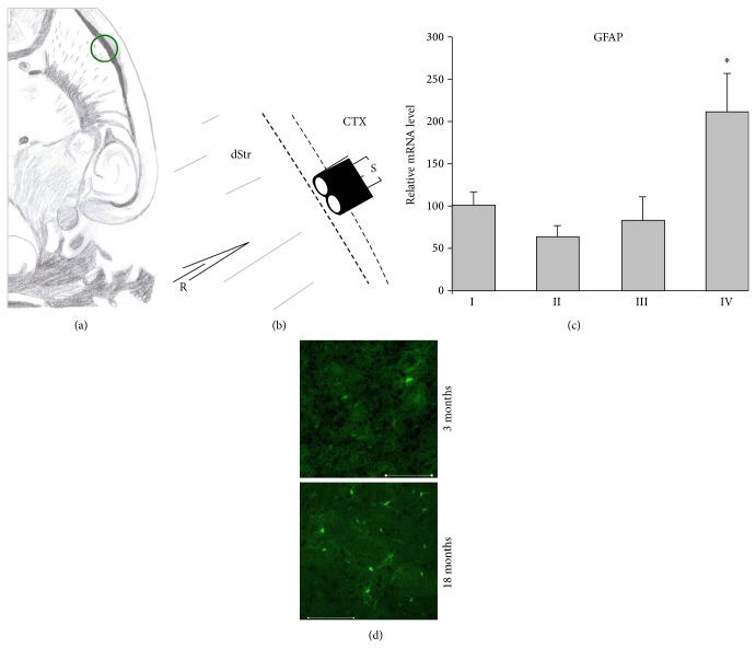
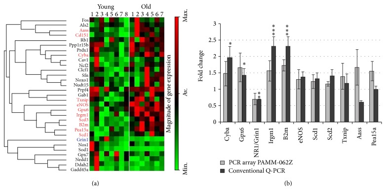
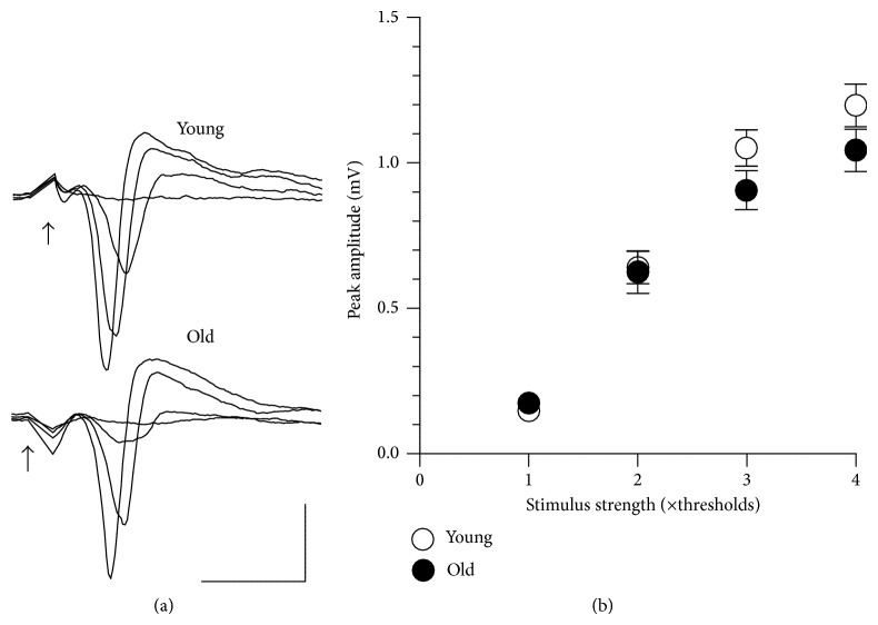
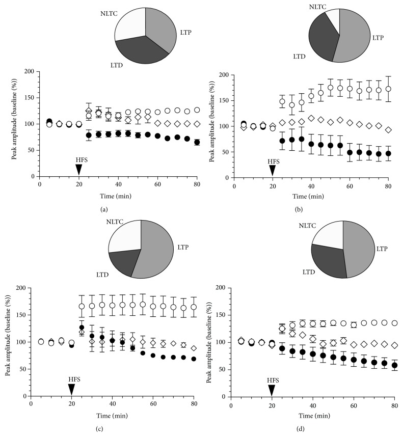
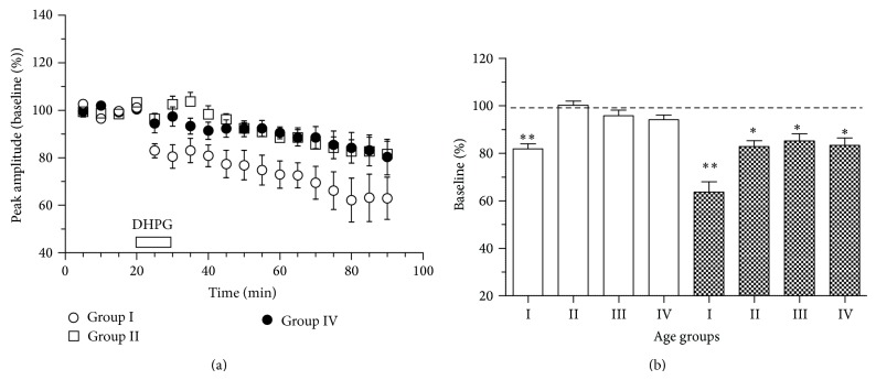
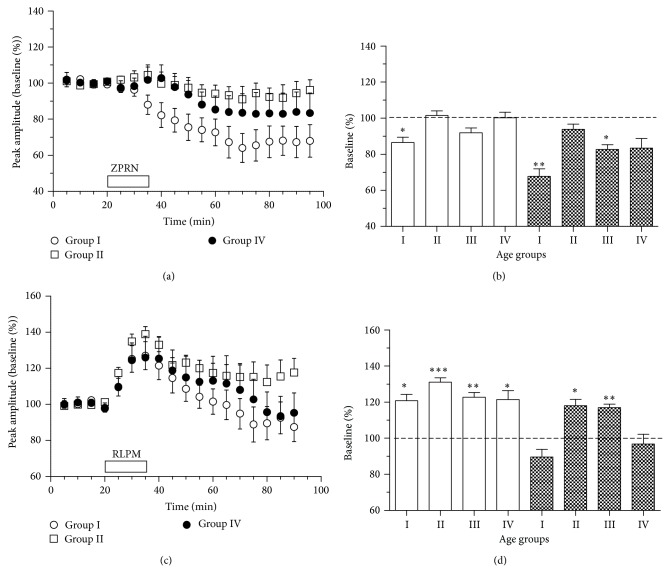
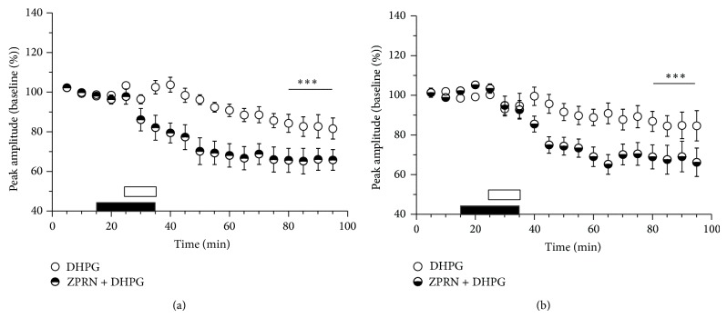
Age-related alterations in the expression of genes and corticostriatal synaptic plasticity were studied in the dorsal striatum of mice of four age groups from young (2-3 months old) to old (18-24 months of age) animals. A significant decrease in transcripts encoding neuronal nitric oxide (NO) synthase and receptors involved in its activation (NR1 subunit of the glutamate NMDA receptor and D1 dopamine receptor) was found in the striatum of old mice using gene array and real-time RT-PCR analysis. The old striatum showed also a significantly higher number of GFAP-expressing astrocytes and an increased expression of astroglial, inflammatory, and oxidative stress markers. Field potential recordings from striatal slices revealed age-related alterations in the magnitude and dynamics of electrically induced long-term depression (LTD) and significant enhancement of electrically induced long-term potentiation in the middle-aged striatum (6-7 and 12-13 months of age). Corticostriatal NO-dependent LTD induced by pharmacological activation of group I metabotropic glutamate receptors underwent significant reduction with aging and could be restored by inhibition of cGMP hydrolysis indicating that its age-related deficit is caused by an altered NO-cGMP signaling cascade. It is suggested that age-related alterations in corticostriatal synaptic plasticity may result from functional alterations in receptor-activated signaling cascades associated with increasing neuroinflammation and a prooxidant state.


Figures

Figure 1
Figure 1
Increased expression of GFAP in the dorsal striatum of old (group IV) mice. (a) Schematic diagram of a horizontal brain slice used for the histological and electrophysiological analysis. The region of counting the GFAP-expressing glial cells and field recording is marked by the green circle. (b) Schematic drawing of relative position of stimulating (S) and recording (R) electrodes in the region marked by the green circle in (a) dStr: dorsal striatum, CTX: cortex. (c) Relative levels of striatal GFAP mRNA in different age groups: group I (n = 8), group II (n = 7), group III (n = 4), and group IV (n = 4). (d) Examples of confocal microscope images with eGFP-GFAP fluorescent cells in striatal slices from a young and an old mouse. Scale bars: 100 µm.
Figure 2
Figure 2
Gene array analysis of striatal transcriptome in young and old mice. (a) Cluster diagram analysis of 31 selected genes expressed in young (n = 8) and old (n = 7) mice. Separation of all samples into 2 age groups was automatically done by the analysis program for only these 31 out of 84 genes included in the PAMM-062Z array. The groups of genes connected by lines on the left of the cluster diagram can be considered coregulated genes. Names of genes whose expression significantly increased with age are written in red. Note an increased expression of genes known to be involved in the immune response (B2m, Irgm1) and oxidative stress (Scd1, Txnip, Gpx6, and Cyba). (b) Validation of gene array data with conventional RT-PCR (primers are given in Supplementary Table 2). Note that significant alterations in the relative mRNA levels for eNOS, Txnip, Aass, and Pea15a revealed by gene array were not confirmed by RT-PCR, whereas transcriptional downregulation with aging for the NMDA receptor NR1 subunit (Grin1) was detected by both assays.
Figure 3
Figure 3
Basal corticostriatal neurotransmission undergoes no significant age-related alterations. (a) Representative examples of corticostriatal field responses recorded in slices from young adult (group I) and old (group IV) mice at n-fold increasing stimulus strength. Each trace represents an average of three responses to the same stimulus voltage. Stimuli are indicated by arrows. Calibrations: vertical: 0.5 mV, horizontal: 5 msec. (b) Averaged stimulus-response plots obtained from corticostriatal preparations from group I (open circles, young, n = 15) and group IV (filled circles, old, n = 18) mice.
Figure 4
Figure 4
Long-term alterations in corticostriatal neurotransmission after high-frequency stimulation of synaptic input in mice of four age groups. The time course (plots) and the incidence (circle diagrams) of long-term potentiation (LTP, open circles), long-term depression (LTD, filled circles), and no long-term changes (NLTC, diamonds) in the striatum of group I (a), group II (b), group III (c), and group IV (d) mice.
Figure 5
Figure 5
Aging weakens depression of corticostriatal responses induced by group I mGluR activation. (a) The mean time course of alterations in corticostriatal field responses induced by the group I mGluR agonist DHPG (100 µM) in striatal slices from mice of different ages (n = 8-9 per group). (b) The magnitudes of initial (averaged through minutes 5–20 after DHPG, open bars) and long-term (averaged through minutes 55–70 after DHPG, filled bars) depression in the striatal slices from mice of four age groups. Significant decrease of corticostriatal responses compared to the baseline is marked by asterisks: * P < 0.05, ** P < 0.01, paired t-test.
Figure 6
Figure 6
Age-dependence of alterations in corticostriatal field responses induced by phosphodiesterase (PDE) inhibitors. (a) The mean time course of alterations in corticostriatal field responses in striatal slices from mice of different ages after exposure to the inhibitor of cGMP-specific PDE5 zaprinast (ZPRN, 40 µM, application period is marked by open bar). (b) Initial (averaged through minutes 10–25 after ZPRN, open bars) and long-term (averaged through minutes 60–75 after ZPRN, filled bars) changes in field response amplitudes induced by ZPRN in four age groups (n = 8 each). (c) The mean time course of alterations in corticostriatal field responses in striatal slices from mice of different ages after exposure to the inhibitor of cAMP-specific PDE4 rolipram (RLPM, 20 µM, marked by bar). (d) Initial (averaged through minutes 5–20 after RLPM, open bars) and long-lasting (averaged through minutes 55–70 after RLPM, filled bars) changes in field response amplitudes induced by RLPM in four age groups (n = 7–9 per group). In (b) and (d), asterisks indicate significant decline of average response amplitude from the baseline, * P < 0.05, ** P < 0.01, and *** P < 0.001, paired t-test.
Figure 7
Figure 7
Age-related reduction of DHPG-LTD is reversed by the phosphodiesterase 5 inhibitor zaprinast (ZPRN). The time course of relative changes in field response amplitudes in corticostriatal slices from group II (a) and group III (b) mice after application of DHPG in the absence (DHPG) or presence of zaprinast (ZPRN + DHPG). Period of DHPG application is marked by open bar; ZPRN was applied 10 min before and together with DHPG (marked by filled bar). Significant differences between the DHPG-LTD magnitudes in the absence and presence of ZPRN are marked by asterisks, *** P < 0.001, t-test.

Similar articles

Cited by

References

    1. Kalpouzos G., Persson J., Nyberg L. Local brain atrophy accounts for functional activity differences in normal aging. Neurobiology of Aging. 2012;33(3):623.e1–623.e13. doi: 10.1016/j.neurobiolaging.2011.02.021. - DOI - PubMed
    1. Langenecker S. A., Briceno E. M., Hamid N. M., Nielson K. A. An evaluation of distinct volumetric and functional MRI contributions toward understanding age and task performance: a study in the basal ganglia. Brain Research. 2007;1135(1):58–68. doi: 10.1016/j.brainres.2006.11.068. - DOI - PMC - PubMed
    1. Marchand W. R., Lee J. N., Suchy Y., et al. Age-related changes of the functional architecture of the cortico-basal ganglia circuitry during motor task execution. NeuroImage. 2011;55(1):194–203. doi: 10.1016/j.neuroimage.2010.12.030. - DOI - PubMed
    1. Calabresi P., Centonze D., Gubellini P., et al. Synaptic transmission in the striatum: from plasticity to neurodegeneration. Progress in Neurobiology. 2000;61(3):231–265. doi: 10.1016/s0301-0082(99)00030-1. - DOI - PubMed
    1. Kreitzer A. C., Malenka R. C. Striatal plasticity and basal ganglia circuit function. Neuron. 2008;60(4):543–554. doi: 10.1016/j.neuron.2008.11.005. - DOI - PMC - PubMed

Publication types

MeSH terms

LinkOut - more resources