. 2015 Mar 5:6:651-664.
doi: 10.3762/bjnano.6.66. eCollection 2015.

Influence of gold, silver and gold-silver alloy nanoparticles on germ cell function and embryo development

Affiliations

Influence of gold, silver and gold-silver alloy nanoparticles on germ cell function and embryo development

Ulrike Taylor et al. Beilstein J Nanotechnol. .

Abstract

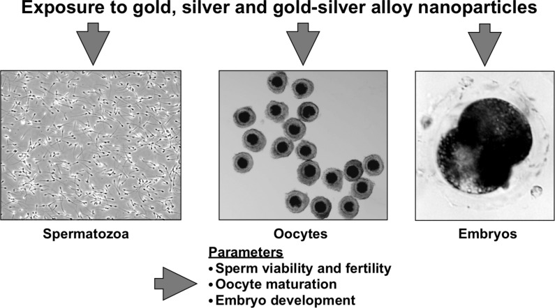
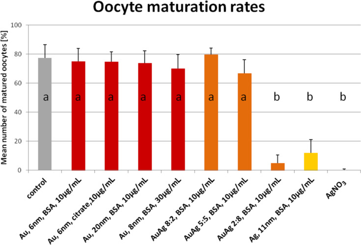
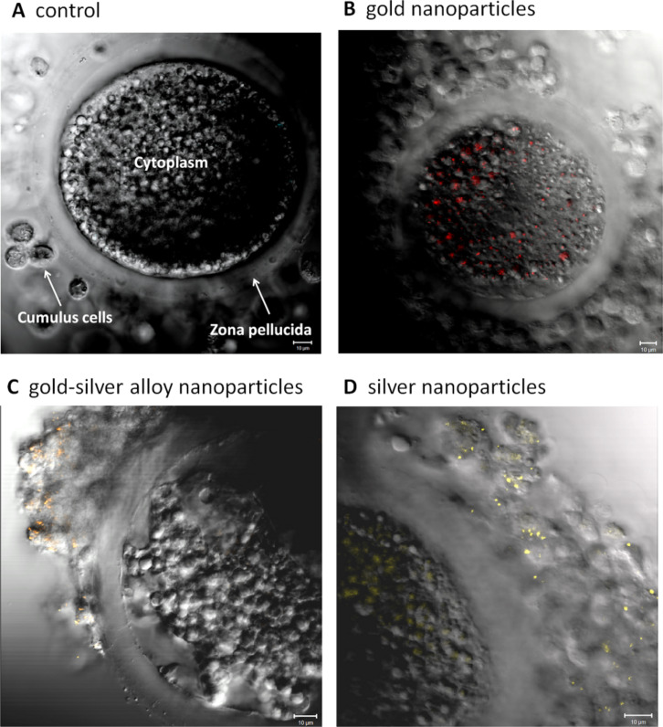
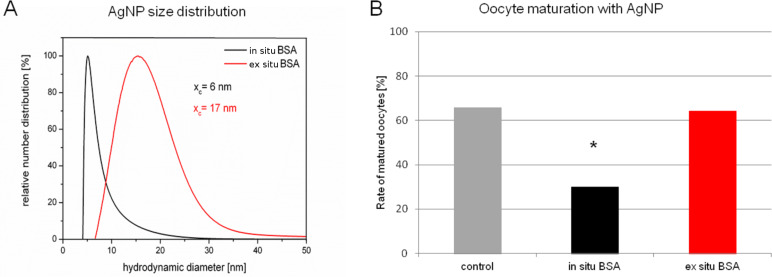
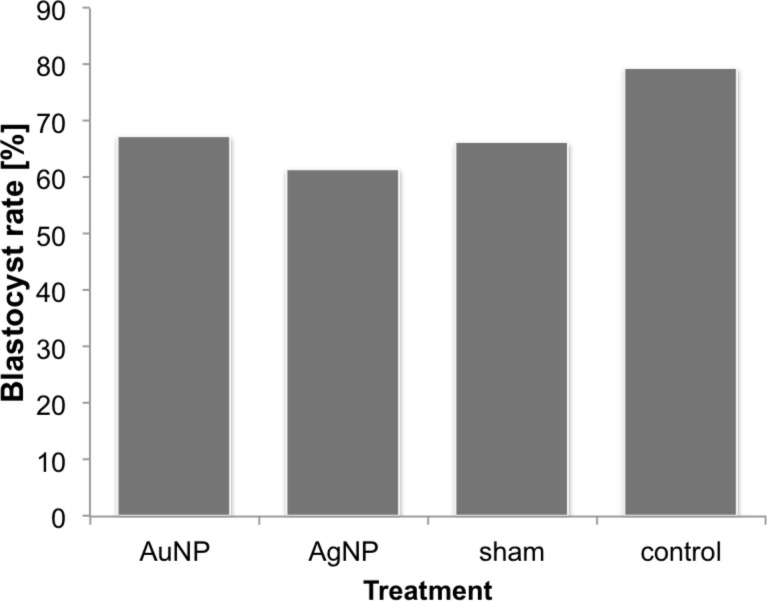
The use of engineered nanoparticles has risen exponentially over the last decade. Applications are manifold and include utilisation in industrial goods as well as medical and consumer products. Gold and silver nanoparticles play an important role in the current increase of nanoparticle usage. However, our understanding concerning possible side effects of this increased exposure to particles, which are frequently in the same size regime as medium sized biomolecules and accessorily possess highly active surfaces, is still incomplete. That particularly applies to reproductive aspects, were defects can be passed onto following generations. This review gives a brief overview of the most recent findings concerning reprotoxicological effects. The here presented data elucidate how composition, size and surface modification of nanoparticles influence viablility and functionality of reproduction relevant cells derived from various animal models. While in vitro cultured embryos displayed no toxic effects after the microinjection of gold and silver nanoparticles, sperm fertility parameters deteriorated after co-incubation with ligand free gold nanoparticles. However, the effect could be alleviated by bio-coating the nanoparticles, which even applies to silver and silver-rich alloy nanoparticles. The most sensitive test system appeared to be in vitro oocyte maturation showing a dose-dependent response towards protein (BSA) coated gold-silver alloy and silver nanoparticles leading up to complete arrest of maturation. Recent biodistribution studies confirmed that nanoparticles gain access to the ovaries and also penetrate the blood-testis and placental barrier. Thus, the design of nanoparticles with increased biosafety is highly relevant for biomedical applications.

Keywords: bimetallic nanoparticles, nano gold; nano silver; ontogenesis, oocyte; reprotoxicity; spermatozoa.


Figures

Figure 1
Figure 1
Schematic representation of experiments conducted within the collaboration project REPROTOX.
Figure 2
Figure 2
(A) Exemplary AuAg colloids with different molar fractions. (B) Correlation of gold molar fraction with the maximum surface plasmon resonance extinction peak. (C) TEM-EDX line scan with inset showing high-angular annular dark field micrograph. (D) TEM micrograph of a Ag50Au50 nanoparticle dispersion after stabilisation with BSA. (E) Aluminium batch chamber for the synthesis of silver and gold–silver alloy nanoparticles. Reproduced with permission from [50]. Copyright 2014 Royal Society of Chemistry.
Figure 3
Figure 3
Representative TEM-micrographs of bovine spermatozoa after co-incubation with gold nanoparticles (AuNP) (10 µg/mL Au) for 2 h at 37 °C. (A) Ligand-free AuNP, (B) oligonucleotide-conjugated AuNP, (C) BSA-coated AuNP. Arrows point out AuNP. Inserts depict the displayed sperm section in total. Above each section the relevant nanoparticle type is displayed schematically. PM = plasma membrane; Ac = acrosome; Nu = nucleus (adapted from [–50]).
Figure 4
Figure 4
Sperm viability parameters after co-incubation of sperm for 2 h at 37 °C with various nanoparticle types and a silver nitrate control. Nanoparticle concentration was 10 µg/mL. (A) Motility assessed with Computer Assissted Sperm Analysis, (B) Membrane integrity assessed with propidium iodide stain and flow cytometer, (C) morphology assessed with phase contrast microscope and evaluation of 200 sperm cells per group per day. Shown are percentage of spermatozoa, which differ compared to the control [values are mean ± SD]. Reproduced with permission from [50]. Copyright 2014 Royal Society of Chemistry.
Figure 5
Figure 5
Oocyte maturation rates after 46 h of in vitro maturation in the presence of various nanoparticle types or silver nitrate in the maturation medium during the complete in vitro maturation time. Maturation rate defined in this case as percentage of oocytes displaying a metaphase plate and extruded polar body (second meiotic division) [values are mean ± SD; a,b p < 0.05]. 350 oocytes were assessed per group. Nanoparticle concentration was 10 µg/mL and all particles were conjugated with bovine serum albumin. Reproduced with permission from [50]. Copyright 2014 Royal Society of Chemistry.
Figure 6
Figure 6
Representative laser scanning microscope images of porcine cumulus–oocyte complexes after 46 h co-incubation during in vitro maturation. (A) Negative control; (B) gold nanoparticles; (C) gold–silver alloy nanoparticles; (D) silver nanoparticles; bars = 10 micrometer. Reproduced with permission from [50]. Copyright 2014 Royal Society of Chemistry.
Figure 7
Figure 7
(A) Number weighted size distribution of AgNP in situ (red line) and ex situ (black line) conjugated to bovine serum albumin (BSA) as measured by disc centrifugation. xc has been calculated by log-normal fitting. (B) Oocyte maturation rates after exposure to AgNP in situ or ex situ bioconjugated to BSA. *p < 0.05.
Figure 8
Figure 8
Blastocyst development rates after microinjection of nanoparticles into 2-cell-stage murine embryos (AuNP-injection, AgNP-injection, sham injection, handling control) (adapted from [51]).

References

    1. Kim S W, Kwak J I, An Y J. Environ Sci Technol. 2013;47:5393–5399. doi: 10.1021/es304511z. - DOI - PubMed
    1. Ramalho-Santos J, Amaral S. Mol Cell Endocrinol. 2013;379:74–84. doi: 10.1016/j.mce.2013.06.005. - DOI - PubMed
    1. Mcevoy T G, Coull G D, Broadbent P J, Hutchinson J S, Speake B K. J Reprod Fertil. 2000;118:163–170. doi: 10.1530/jrf.0.1180163. - DOI - PubMed
    1. Parks J E, Lynch D V. Cryobiology. 1992;29:255–266. doi: 10.1016/0011-2240(92)90024-V. - DOI - PubMed
    1. Flesch F M, Gadella B M. Biochim Biophys Acta. 2000;1469:197–235. doi: 10.1016/S0304-4157(00)00018-6. - DOI - PubMed

LinkOut - more resources