. 2015:2015:127828.
doi: 10.1155/2015/127828. Epub 2015 Mar 2.

Inhibitory and apoptosis-inducing effects of Newcastle disease virus strain AF2240 on mammary carcinoma cell line

Affiliations

Inhibitory and apoptosis-inducing effects of Newcastle disease virus strain AF2240 on mammary carcinoma cell line

Umar Ahmad et al. Biomed Res Int. 2015.

Abstract

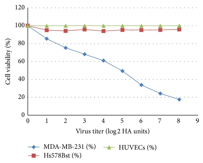
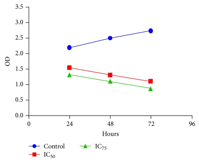
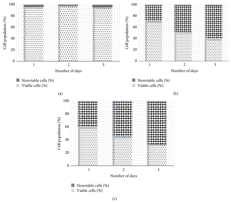
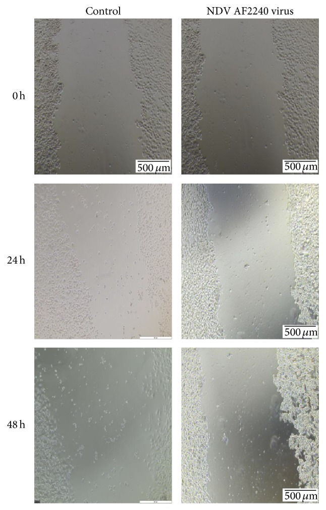
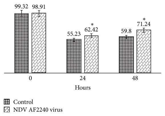
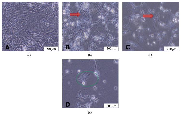
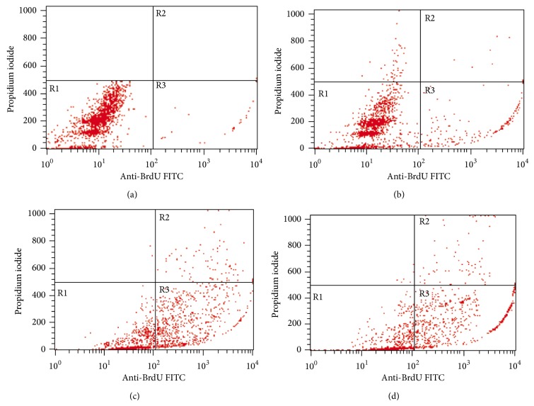
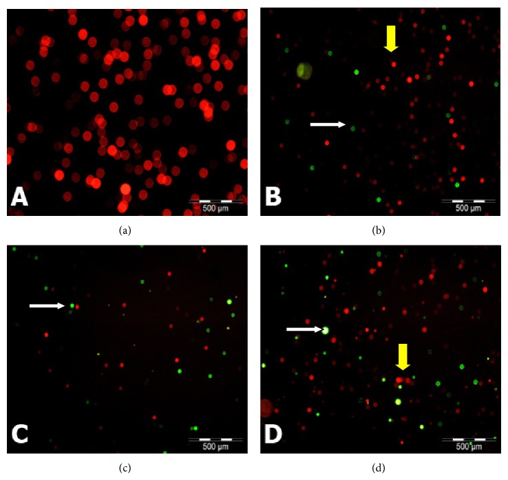
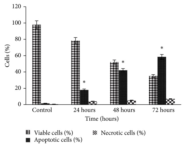
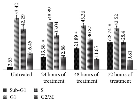
Breast cancer is the malignant tumour that developed from cells of the breast and is the first leading cause of cancer death among women worldwide. Surgery, radiotherapy, and chemotherapy are the available treatments for breast cancer, but these were reported to have side effects. Newcastle disease virus (NDV) known as Avian paramyxovirus type-1 (APMV1) belongs to the genus Avulavirus in a family Paramyxoviridae. NDV is shown to be a promising anticancer agent, killing tumour cells while sparing normal cells unharmed. In this study, the oncolytic and cytotoxic activities of NDV AF2240 strain were evaluated on MDA-MB-231, human mammary carcinoma cell line, using MTT assay, and its inhibitory effects were further studied using proliferation and migration assays. Morphological and apoptotic-inducing effects of NDV on MD-MB-231 cells were observed using phase contrast and fluorescence microscopes. Detection of DNA fragmentation was done following terminal deoxyribonucleotide transferase-mediated Br-dUTP nick end labeling staining (TUNEL) assay, which confirmed that the mode of death was through apoptosis and was quantified by flow cytometry. Furthermore, analysis of cellular DNA content demonstrated that the virus caused an increase in the sub-G1 phase (apoptotic peak) of the cell cycle. It appears that NDV AF2240 strain is a potent anticancer agent that induced apoptosis in time-dependent manner.


Figures

Figure 1
Figure 1
Scatter plot of the MTT Assay with regression line of NDV AF2240 IC50 (24.7 HAU/mL) values determined in MDA-MB-231 cell lines (y = −10.251x + 98.206) after 72 hours of treatment. This result indicated the significant inhibitory effect of the virus on MD-MB-231, but no effect was observed on Hs578Bst and HUVEC cells. Data shown are mean ± SD of triplicate (n = 3) independent experiments. Correlation coefficient R 2 = 0.99176.
Figure 2
Figure 2
Quick Cell Proliferation Colorimetric Assay for IC50 and IC75 concentration values of the NDV AF2240 virus strain against MDA-MB-231 cells at 24, 48, and 72 hours after treatment. As indicated, the growth rates for the treated cells decreased significantly as compared with the control cells (P < 0.05). However, no significant different (P > 0.05) was observed between the two-virus concentrations.
Figure 3
Figure 3
Percentage of viable and nonviable cells for MDA-MB-231 cell line exposed to time course. (a) Untreated (cancer) control cells, (b) cells treated with IC50 concentration value (25.99 HAU/mL) of NDV AF2240, and (c) cells treated with IC75 concentration (128 HAU/mL) of NDV AF2240. Results were expressed as mean (%) from triplicate experiments.
Figure 4
Figure 4
Effect of NDV AF2240 strain on cell migration by scratch wound assay. Confluent monolayers were cultured without (control) and with IC50 (25.99 HAU/mL) value of the virus and the migration was evaluated by wound healing assay at 24 and 48 h. Cells were photographs at 0, 24, and 48 hours after viral treatment. Note: at 48-hour time, the virus ruptured the cells and disjointed them from neighboring cells.
Figure 5
Figure 5
Percentage open wound area performed with TScratch software. Data are shown as % of open wound area in comparison with control cells. Cells were treated with IC50 (25.99 HAU/mL) value of NDV AF2240 at 0 hour (99.32%), and the results were expressed as mean ± SD from triplicate (n = 3) independent experiments. A significant different (* P < 0.05) was observed when the control cells were compared with the treated cells.
Figure 6
Figure 6
Phase contrast micrographs of MDA-MB-231 breast cancer cell lines treated with NDV AF2240 IC50 concentration value. (a) Untreated MDA-MB-231 cell line after (b) 24 hours (c) 48 hours, and (d) 72 hours of treatment. The virus caused the cells to lose contact with adjacent cells (red arrow) and cell membrane disruption (green circle). Mag 20x.
Figure 7
Figure 7
Contour diagram of BrdUTP/PI flow cytometry for MDA-MB-231 breast cancer cell lines treated with NDV AF2240 IC50 concentration value. (a) Untreated cells after (b) 24 hours (c) 48 hours, and (d) 72 hours of treatment. The lower left quadrants of each panel (R1) show the viable cells, which exclude PI and are negative for BrdUTP binding. The upper right quadrants (R2) contain the nonviable, necrotic cells, positive for BrdUTP binding and for PI uptake. The lower right quadrants (R3) represent the fragmented DNA (apoptotic cells), BrdUTP positive, and PI negative.
Figure 8
Figure 8
Fluorescent micrographs of MDA-MB-231 breast cancer cells treated with NDV AF2240 stained by TUNEL assay technique (Apo-BrdU), double stained with fluorescein Br-dUTP and PI/RNase A solution: (a) untreated (cancer control) cells after (b) 24 hours (c) 48 hours, and (d) 72 hours of treatment periods. Viable cells show orange-red nuclei in color (yellow arrow), whereas apoptotic cells show yellow to greenish nuclei (white arrow). Mag 20x.
Figure 9
Figure 9
Fluorescent microscopy examinations. Percentages of viable, apoptotic, and necrotic cells in MDA-MB-231 cell population treated with NDV AF2240 after 24, 48, and 72 hours. Data shown are mean ± SD from triplicate (n = 3) independent experiments. Cells death via apoptosis increased significantly (* P < 0.05) in time-dependent manner. However, no significant (P > 0.05) difference was observed in the necrotic cells.
Figure 10
Figure 10
Flow cytometric cell cycle analysis of MDA-MB-231 cell after staining with BrdUTP/PI. Cells treated with NDV AF2240 strain at IC50. Data shown in mean ± SD from triplicate (n = 3) independent experiments. One-way ANOVA analysis was run with Tukey's HSD Post Hoc multiple comparison tests. Significant at (* P < 0.05) compared with the control.

References

    1. McPherson K., Steel C. M., Dixon J. M. ABC of breast diseases: breast cancer—epidemiology, risk factors, and genetics. British Medical Journal. 2000;321(7261):624–628. doi: 10.1136/bmj.321.7261.624. - DOI - PMC - PubMed
    1. Burke W., Daly M., Garber J., et al. Recommendations for follow-up care of individuals with an inherited predisposition to cancer: II. BRCA1 and BRCA2. The Journal of the American Medical Association. 1997;277(12):997–1003. doi: 10.1001/jama.1997.03540360065034. - DOI - PubMed
    1. Loman N., Johannsson O., Kristoffersson U., Olsson H., Borg Å. Family history of breast and ovarian cancers and BRCA1 and BRCA2 mutations in a population-based series of early-onset breast cancer. Journal of the National Cancer Institute. 2001;93(16):1215–1223. doi: 10.1093/jnci/93.16.1215. - DOI - PubMed
    1. Goldhirsch A., Wood W. C., Gelber R. D., Coates A. S., Thürlimann B., Senn H.-J. Meeting highlights: updated international expert consensus on the primary therapy of early breast cancer. Journal of Clinical Oncology. 2003;21(17):3357–3365. doi: 10.1200/jco.2003.04.576. - DOI - PubMed
    1. You L., He B., Xu Z., McCormick F., Jablons D. M. Future directions: uncolytic viruses. Clinical Lung Cancer. 2004;5(4):226–230. doi: 10.3816/clc.2004.n.003. - DOI - PubMed

Publication types

LinkOut - more resources