. 2015 Jun;21(6):1282-96.
doi: 10.1097/MIB.0000000000000371.

Downregulation of FoxC2 Increased Susceptibility to Experimental Colitis: Influence of Lymphatic Drainage Function?

Affiliations

Downregulation of FoxC2 Increased Susceptibility to Experimental Colitis: Influence of Lymphatic Drainage Function?

Felix Becker et al. Inflamm Bowel Dis. 2015 Jun.

Abstract

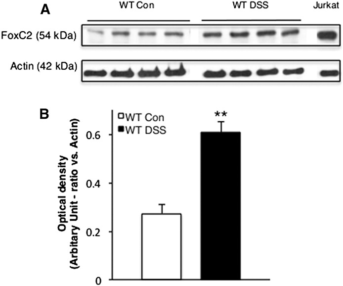
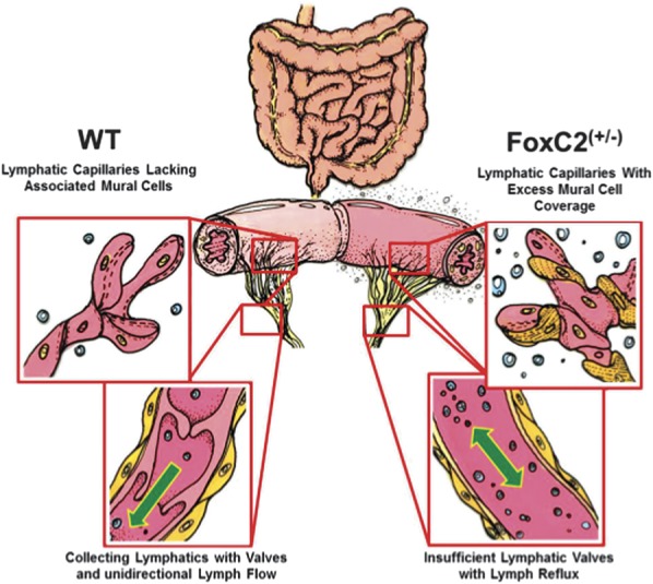
Background: Although inflammation-induced expansion of the intestinal lymphatic vasculature (lymphangiogenesis) is known to be a crucial event in limiting inflammatory processes, through clearance of interstitial fluid and immune cells, considerably less is known about the impact of an impaired lymphatic clearance function (as seen in inflammatory bowel diseases) on this cascade. We aimed to investigate whether the impaired intestinal lymphatic drainage function observed in FoxC2 mice would influence the course of disease in a model of experimental colitis.

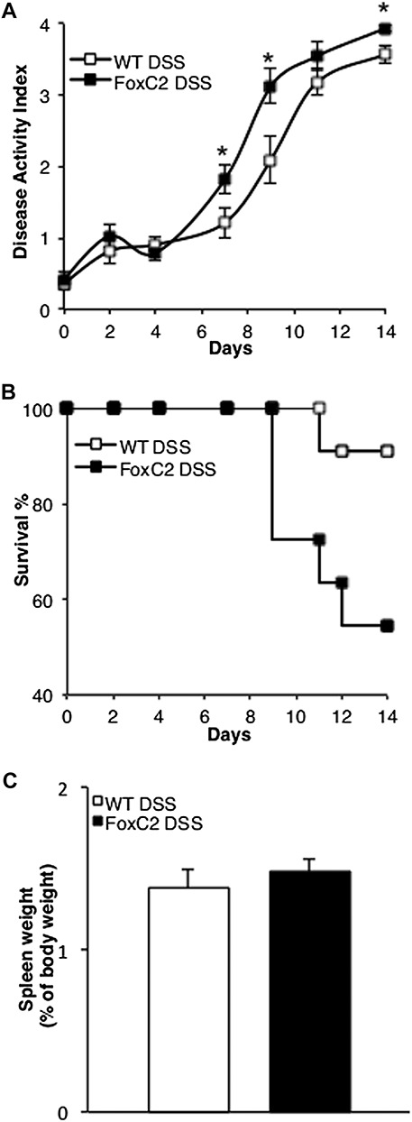
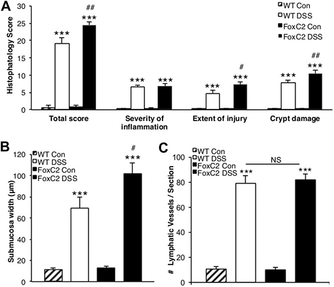
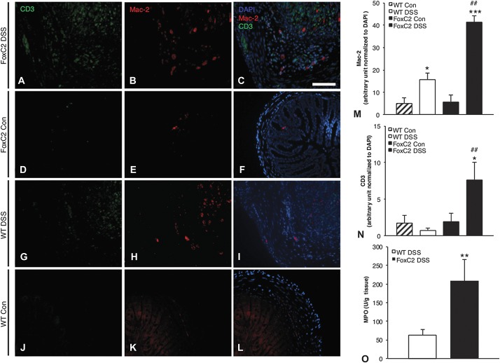
Methods: Acute dextran sodium sulfate colitis was induced in wild-type and haploinsufficient FoxC2 mice, and survival, disease activity, colonic histopathological injury, neutrophil, T-cell, and macrophage infiltration were evaluated. Functional and structural changes in the intestinal lymphatic vessel network were analyzed, including submucosal edema, vessel morphology, and lymphatic vessel density.

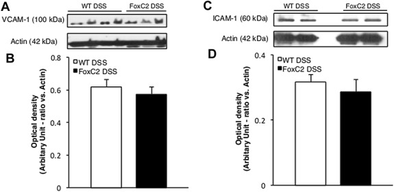
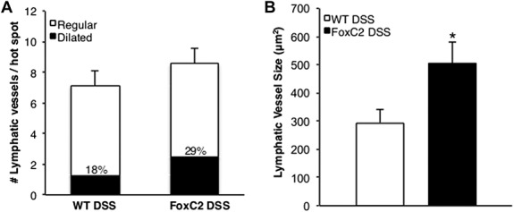
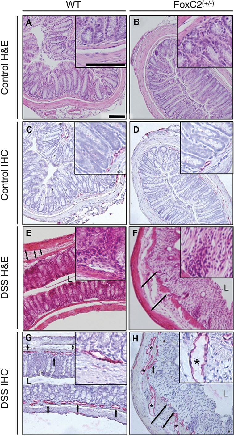
Results: We found that FoxC2 downregulation in FoxC2 mice significantly increased the severity and susceptibility to experimental colitis, as displayed by lower survival rates, increased disease activity, greater histopathological injury, and elevated colonic neutrophil, T-cell, and macrophage infiltration. These findings were accompanied by structural (dilated torturous lymphatic vessels) and functional (greater submucosal edema, higher immune cell burden) changes in the intestinal lymphatic vasculature.

Conclusions: These results indicate that sufficient lymphatic clearance plays a crucial role in limiting the initiation and perpetuation of experimental colitis and those disturbances in the integrity of the intestinal lymphatic vessel network could intensify intestinal inflammation. Future therapies might be able to exploit these processes to restore and maintain adequate lymphatic clearance function in inflammatory bowel disease.


Conflict of interest statement

J. S. Alexander is currently receiving a grant (W81XWH-11-1-0577, Lymphatic Vascular-based Therapy in IBD) from the Department of Defense. I. Tsunoda is supported by the National Institute of General Sciences COBRE Grant (P30-GM110703). F. Becker is supported by a fellowship grant from Abbvie Corporation (16,165) and the German Research Foundation (DFG, BE 5619/1-1). The remaining authors have no conflicts of interest to disclose.

Figures

FIGURE 1
FIGURE 1
FoxC2 protein expression was significantly increased during acute DSS-induced experimental colitis. A, Representative Western blot analysis of FoxC2 (54 kDa) and actin (42 kDa) expression in colon lysates (30 μg total protein) from control and DSS-treated WT mice. Jurkat cell lysate (10 μg total protein) was used as positive control. B, Densitometric analysis for FoxC2 optical density in ratio to actin (n = 4, Student's t test, **P < 0.001 versus WT Control).
FIGURE 2
FIGURE 2
FoxC2(+/−) mice challenged with 2% DSS exhibited a higher mortality and greater disease activity but a similar systemic inflammatory response compared with DSS-treated WT mice. A, DSS-treated FoxC2(+/−) mice showed a 45% mortality rate compared with 9% mortality in DSS-treated WT mice. Kaplan–Meier curves display changes from initial n = 11 in both groups over the 14 days course of DSS treatment. B, Disease activity (consisting of the parameters stool blood, stool form, and weight loss) in FoxC2(+/−) DSS-treated mice was significantly elevated on days 7, 9, and 14 (*P < 0.05) compared with WT DSS (n = 11, Student's t test). Disease activity in both groups differed significantly (not shown) from control starting on day 4 (P < 0.01, repeated measures analysis of variance, with Dunnett's post hoc testing). C, Spleen weights (measured [in grams] and expressed as percentage of the body weight [in grams] at day 14) were analyzed as an index of systemic inflammation. DSS-treated WT and DSS-treated FoxC2(+/−) mice showed similar spleen weights after 14 days of DSS colitis (n = 5 to 7).
FIGURE 3
FIGURE 3
FoxC2(+/−) mice developed greater histopathological injury and submucosal edema in DSS-induced colitis, while exhibited normal inflammation-induced lymphangiogenesis (A) total histopathology score (consisting of the individual parameters severity of inflammation, extent of injury, and crypt damage) was significantly greater in DSS-treated WT and DSS-treated FoxC2(+/−) mice compared with untreated controls. When colitis groups were compared, FoxC2(+/−) mice showed an elevated total score and an increase in the individual parameters crypt damage and extent of injury. B, Submucosa edema (defined as the range between tunica mucosa and muscularis in microns) was significantly greater after DSS-induction in WT and FoxC2(+/−) mice compared with untreated controls. When comparing DSS-treated WT with DSS-treated FoxC2(+/−) mice, the submucosal edema in FoxC2(+/−) mice was significantly increased. C, LVD (number of lymphatic vessels per section) was determined as a marker for inflammation-induced lymphangiogenesis in LYVE-1–stained colonic cross-sections for untreated control and DSS-treated WT and FoxC2(+/−) mice. Compared with controls, the induction of DSS colitis provoked a significant increase in LVD in FoxC2(+/−) and WT mice, whereas no differences were seen between the colitis groups (n = 5 to 7), one-way analysis of variance, Bonferroni posttesting. ***P < 0.001 versus untreated control; ##P < 0.01 versus WT DSS, #P < 0.05 versus WT DSS; NS = not significant.
FIGURE 4
FIGURE 4
FoxC2(+/−) mice showed significantly increased intestinal neutrophil, macrophage, and T-cell infiltration compared with WT mice in DSS colitis (A–L). Macrophages and T cells in the colonic submucosa of untreated WT (J–L) and FoxC2(+/−) (D–F) as well as DSS-treated WT (G–I) and DSS-treated FoxC2(+/−) mice (A–C) were quantified using immunofluorescent staining against Mac-2 (macrophages; B, E, H, K) and CD3 (T cells; A, D, G, J). To better visualize the presence of these immune cells, representative single color and merged pictures with DAPI (C, F, I, L) staining are shown. To quantify the colonic Mac-2+ macrophage and CD3+ T-cell content, Mac-2 (M) and CD3 (N) signals within the submucosa were normalized to the area of submucosal DAPI signal (expressed in arbitrary units) (×200 magnification, scale bar 100 μm, n = 5 to 7, one-way analysis of variance, Bonferroni posttesting). *P < 0.05 versus untreated control, ***P < 0.001 versus untreated control; ##P < 0.01 versus WT DSS. (O) MPO activity (in Units per gram tissue) was measured as an index for tissue neutrophil content. DSS-treated FoxC2(+/−) mice showed a significant increase in colonic MPO activity compared with DSS-treated WT mice (n = 5 to 7, Student's t test; **P < 0.01 versus WT DSS).
FIGURE 5
FIGURE 5
ICAM-1 and VCAM-1 protein expression was not altered during DSS colitis upon FoxC2 insufficiency. Representative Western blot analysis of VCAM-1 (A, ∼100 kDa), ICAM-1 (C, ∼60 kDa), and actin (∼42 kDa) expression in colon lysates (30 μg total protein) from DSS-treated WT and DSS-treated FoxC2(+/−) mice. Densitometric analysis for VCAM-1 (B) and ICAM-1 (D) optical density in ratio to actin (n = 4).
FIGURE 6
FIGURE 6
DSS-treated FoxC2(+/−) mice exhibited dilated lymphatic vessels compared with DSS-treated WT mice. In LYVE-1–stained colonic cross-sections, 3 areas (“hot spots”) of the highest LVD were selected in which the number of lymphatic vessels per hot spot was counted and the lymphatic vessel size (expressed as vessel lumen area in square microns) was measured. Dilated lymphatic vessels were defined as those vessels whose mean vessel area was greater than 2 SDs above those in untreated control mice, whereas regular lymphatic vessels were defined as those vessels whose mean vessel area was within 2 SDs of those in untreated control mice. A, Compared with DSS-treated WT mice, DSS-treated FoxC2(+/−) mice showed a similar number of lymphatic vessels per hot spot, of which 18% ± 5.5% in WT and 29% ± 7.8% in FoxC2(+/−) mice were defined as “dilated.” B, Compared with DSS-treated WT mice, DSS-treated FoxC2(+/−) mice developed more numerous dilated lymphatic vessels (n = 5 to 7, Student's t test, *P < 0.05 versus WT DSS).
FIGURE 7
FIGURE 7
FoxC2(+/−) mice exhibited dilated and torturous lymphatic vessels and greater submucosal edema (defined as the range between tunica mucosa and muscularis) and histopathological injury. Representative images of colonic cross-sections from control and DSS-treated WT (left) and control and DSS-treated FoxC2(+/−) mice (right); hematoxylin–eosin (H&E, blue: nuclei; red: cytoplasm) and immunohistochemistry-stained (IHC, red: LYVE-1–positive lymphatic vessels, blue: hematoxylin-counterstained nuclei), A/C, B/D, E/G, and F/H represent consecutive slides of the same area (L = Lumen, ×160 magnification, inserts ×400 magnification, scale bars 100 μm). A and B, Untreated control mice showed no histological evidence of colitis, whereas the induction of DSS colitis proved typical histopathological changes (E, F) including destruction of the epithelial architecture, crypt damage, cellular inflammation in all colonic layers, and submucosal edema. These morphological changes were increased in DSS-treated FoxC2(+/−) mice (F). DSS-treated WT (G) and FoxC2(+/−) (H) mice showed significant inflammation-induced lymphangiogenesis, accompanied by distinct changes in the lymphatic vessel architecture (black asterisk: dilated lymphatic vessel, black arrows: lymphatic vessels, black double arrows: submucosal width), which were nearly absent in untreated FoxC2(+/−) (D) and WT (C) mice. FoxC2(+/−) developed an increase in submucosal edema and displayed more dilated lymphatic vessels (F and H).
FIGURE 8
FIGURE 8
Intestinal lymphatic networks in WT and haploinsufficient FoxC2(+/−) mice. Left: Normal lymphatic vessel anatomy in WT mice: Blind-ended initial lymphatic capillaries lack associated mural cells (pericytes, SMCs) allowing the penetration of cells and interstitial fluid into the lymphatic lumen. Subsequent larger collecting vessels exhibit an SMC layer and valves maintaining unidirectional lymph flow during regular contraction. Right: Disturbed lymphatic networks in FoxC2(+/−) mice characterized by increased mural cell investment of lymphatic capillaries and valve agenesis in collecting lymphatic vessels. This phenotype was reported to exhibit lymph backflow in the intestinal wall and decreased capacity for lymphatic clearance. A possible functional pathomechanism is most likely the accumulation of (1) impaired permeability in lymphatic capillaries, (2) uneven lumen size, (3) uncoordinated contraction of the aberrant mural SMCs, and (4) lymph backflow because of the valve insufficiency. Challenged with 2% DSS, FoxC2(+/−) mice developed signs of lymphatic clearance failure, characterized by dilated lymphatic vessels, increased tissue edema, and elevated inflammatory cell burden.

Similar articles

Cited by

References

    1. Kappelman MD, Moore KR, Allen JK, et al. Recent trends in the prevalence of Crohn's disease and ulcerative colitis in a commercially insured US population. Dig Dis Sci. 2013;58:519–525. - PMC - PubMed
    1. Burisch J, Jess T, Martinato M, et al. The burden of inflammatory bowel disease in Europe. J Crohns Colitis. 2013;7:322–337. - PubMed
    1. Baumgart DC, Carding SR. Inflammatory bowel disease: cause and immunobiology. Lancet. 2007;369:1627–1640. - PubMed
    1. Baumgart DC, Sandborn WJ. Inflammatory bowel disease: clinical aspects and established and evolving therapies. Lancet. 2007;369:1641–1657. - PubMed
    1. Chidlow JH, Jr, Shukla D, Grisham MB, et al. Pathogenic angiogenesis in IBD and experimental colitis: new ideas and therapeutic avenues. Am J Physiol Gastrointest Liver Physiol. 2007;293:G5–G18. - PubMed

Publication types

MeSH terms

Substances