. 2015 May;89(10):5238-49.
doi: 10.1128/JVI.00395-15. Epub 2015 Mar 11.

Multiple genome segments determine virulence of bluetongue virus serotype 8

Affiliations

Multiple genome segments determine virulence of bluetongue virus serotype 8

Anna Janowicz et al. J Virol. 2015 May.

Abstract

Bluetongue virus (BTV) causes bluetongue, a major hemorrhagic disease of ruminants. In order to investigate the molecular determinants of BTV virulence, we used a BTV8 strain minimally passaged in tissue culture (termed BTV8L in this study) and a derivative strain passaged extensively in tissue culture (BTV8H) in in vitro and in vivo studies. BTV8L was pathogenic in both IFNAR(-/-) mice and in sheep, while BTV8H was attenuated in both species. To identify genetic changes which led to BTV8H attenuation, we generated 34 reassortants between BTV8L and BTV8H. We found that partial attenuation of BTV8L in IFNAR(-/-) mice was achieved by simply replacing genomic segment 2 (Seg2, encoding VP2) or Seg10 (encoding NS3) with the BTV8H homologous segments. Fully attenuated viruses required at least two genome segments from BTV8H, including Seg2 with either Seg1 (encoding VP1), Seg6 (encoding VP6 and NS4), or Seg10 (encoding NS3). Conversely, full reversion of virulence of BTV8H required at least five genomic segments of BTV8L. We also demonstrated that BTV8H acquired an increased affinity for glycosaminoglycan receptors during passaging in cell culture due to mutations in its VP2 protein. Replication of BTV8H was relatively poor in interferon (IFN)-competent primary ovine endothelial cells compared to replication of BTV8L, and this phenotype was determined by several viral genomic segments, including Seg4 and Seg9. This study demonstrated that multiple viral proteins contribute to BTV8 virulence. VP2 and NS3 are primary determinants of BTV pathogenesis, but VP1, VP5, VP4, VP6, and VP7 also contribute to virulence.

Importance: Bluetongue is one of the major infectious diseases of ruminants, and it is listed as a notifiable disease by the World Organization for Animal Health (OIE). The clinical outcome of BTV infection varies considerably and depends on environmental and host- and virus-specific factors. Over the years, BTV serotypes/strains with various degrees of virulence (including nonpathogenic strains) have been described in different geographical locations. However, no data are available to correlate the BTV genotype to virulence. This study shows that BTV virulence is determined by different viral genomic segments. The data obtained will help to characterize thoroughly the pathogenesis of bluetongue. The possibility to determine the pathogenicity of virus isolates on the basis of their genome sequences will help in the design of control strategies that fit the risk posed by new emerging BTV strains.


Figures

FIG 1
FIG 1
In vivo and in vitro phenotype of minimally passaged BTV8L and tissue culture-adapted BTV8H. (A and B) BTV8L and BTV8H replication kinetics in ovine CPT-Tert cells (A) and primary ovine endothelial cells (OvEC) (B). Cells were infected with BTV8L or BTV8H at an MOI of 0.01. Supernatants were collected at 2, 24, 48, and 72 h p.i. and then titrated in BSR cells by limiting dilution analysis. Virus titers are expressed as log10(TCID50/ml). (C) Survival plot of IFNAR−/− mice (n = 5 per group) infected with 300 PFU of BTV8L and BTV8H or mock infected. (D to F) Virulence of BTV8L and BTV8H in sheep. Clinical scores (D), viremia (E), and neutralizing antibodies (F) at 28 days p.i. were measured in infected and mock-infected sheep (n = 5 per group) as described in Materials and Methods. PD50, 50% protective dose.
FIG 2
FIG 2
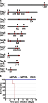
Genetic differences between BTV8L and BTV8H and virulence of rescued viruses in IFNAR−/− mice. (A) Schematic representation of the 10 genomic segments of BTV8L and BTV8H. Mutations in BTV8H compared to the sequence of the minimally passaged BTV8L are indicated with red dots. Nonsynonymous mutations are marked with asterisks, and the numbers relative to the mutated amino acid residues in the corresponding viral proteins are shown. The plus sign indicates a nucleotide insertion. The length of the schematic genome segments and the relative position of mutations are indicative only. (B) Survival plots of IFNAR−/− mice (n = 5 per group) infected intraperitoneally with 300 PFU of rgBTV8L or rgBTV8H.
FIG 3
FIG 3
Virulence of BTV8L/BTV8H reassortants in IFNAR−/− mice. Multiple reassortant viruses were rescued using reverse genetics as described in Materials and Methods. Mortality of IFNAR−/− mice (n = 5 per group) inoculated intraperitoneally with 300 or 3,000 PFU of individual reassortants within the BTV8L or the BTV8H backbone is shown. Note that the combination of segments that resulted in rescue of fully attenuated reassortants is shown in blue boxes. The combination of at least five segments of BTV8L was required to recapitulate the fully virulent phenotype (shown in red boxes).
FIG 4
FIG 4
In vitro replication properties of BTV8L/BTV8H monoreassortants. (A) Growth curves of parental rgBTV8L, rgBTV8H, and monoreassortants containing the BTV8L backbone (gray circle) in CPT-Tert cells. Monolayers were infected with the indicated viruses at an MOI of 0.01, and supernatants were collected at 2, 24, 48, and 72 h p.i. Viral titers were determined by endpoint dilutions. All reassortants, with exception of BTV8L+S2H, showed replication kinetics similar to those of the parental rgBTV8L. (B) Plaques produced in CPT-Tert cells by parental rgBTV8L and rgBTV8H and derived monoreassortants at 48 h p.i. Note the increased plaque size in rgBTV8H and BTV8L+S2H.
FIG 5
FIG 5
Segment 2 of rgBTV8H favors replication in cells expressing glycosaminoglycans. (A) Viral titers produced in CHO and pgsA-745 cells infected with rgBTV8L, rgBTV8H, and reassortant viruses (MOI of 0.01). Supernatants were collected at 72 h p.i. and titrated in BSR cells by limiting dilution analysis. Mean values from three experiments performed in duplicate are shown (error bars correspond to standard deviations). Note that significant differences were observed between titers produced in CHO and pgsA-745 cells by BTV8L+S2H and BTV8H+S2L (***, P < 0.001; two-way analysis of variance followed by a Bonferroni test to identify interactions). (B) Cytopathic effect in CHO and pgsA-745 cells infected with rgBTV8L, rgBTV8H, and reassortant viruses (MOI of 0.01). Cells were stained with crystal violet at 72 h p.i.
FIG 6
FIG 6
Replication kinetics of parental and monoreassortant viruses in primary OvEC. OvEC were infected at an MOI of 0.01 with rgBTV8L, rgBTV8H, and monoreassortants containing the BTV8L backbone (gray circle). Viral titers were determined at the specified time points by limiting dilution assays. Each panel shows growth curves of both parental viruses and a specific monoreassortant (as labeled).
FIG 7
FIG 7
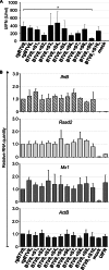
IFN production and gene expression induced by infection of OvEC by rgBTV8L, rgBTV8H, and BTV8L/BTV8H monoreassortants. OvEC were infected with rgBTV8L, rgBTV8H, and monoreassortants within the BTV8L backbone (MOI of 1). (A) IFN protection assay. Supernatants were collected at 18 h p.i., inactivated by UV treatment, and used in a biological assay to estimate the amount of IFN present, as described in Materials and Methods. The only major differences were observed in cells infected with BTV8L+S9H, where the amount of IFN released was significantly lower than what was found in cells infected with rgBTV8L (P < 0.05; one-way analysis of variance followed by Dunnett's multiple-comparison test to dissect individual interactions). (B) IFN-β, ActB, RSAD2, and Mx1 gene expression. mRNA was measured by qPCR in OvEC at 18 h p.i. with parental and reassortant viruses (MOI of 1) as described in Materials and Methods. Mock-treated and UIFN-treated cells were used as controls. Panels show gene expression relative to rgBTV8L and normalized to GAPDH gene levels.
FIG 8
FIG 8
Replication of rgBTV8L, rgBTV8H, and BTV8L/BTV8H monoreassortants in CPT-Tert cells pretreated with universal IFN (UIFN). Viral titers produced in untreated and IFN-pretreated CPT-Tert cells by parental and reassortant viruses at 48 h p.i. at an MOI of 0.01. Mean values from three experiments performed in duplicate are shown (error bars correspond to standard deviations). *, P < 0.05; **, P < 0.01; ***, P < 0.001 (one-way analysis of variance followed by Dunnett's multiple-comparison test to dissect individual interactions).

Similar articles

Cited by

References

    1. Price DA, Hardy WT. 1954. Isolation of the bluetongue virus from Texas sheep-Culicoides shown to be a vector. J Am Vet Med Assoc 124:255–258. - PubMed
    1. MacLachlan NJ, Drew CP, Darpel KE, Worwa G. 2009. The pathology and pathogenesis of bluetongue. J Comp Pathol 141:1–16. doi:10.1016/j.jcpa.2009.04.003. - DOI - PubMed
    1. Mellor PS, Baylis M, Mertens PP. 2009. Bluetongue. Academic Press, London, United Kingdom.
    1. Roy P, Noad R. 2006. Bluetongue virus assembly and morphogenesis. Curr Top Microbiol Immunol 309:87–116. doi:10.1007/3-540-30773-7_4. - DOI - PubMed
    1. Ratinier M, Caporale M, Golder M, Franzoni G, Allan K, Nunes SF, Armezzani A, Bayoumy A, Rixon F, Shaw A, Palmarini M. 2011. Identification and characterization of a novel non-structural protein of bluetongue virus. PLoS Pathog 7:e1002477. doi:10.1371/journal.ppat.1002477. - DOI - PMC - PubMed

Publication types

MeSH terms

LinkOut - more resources