. 2015 Mar 30;10(3):e0122839.
doi: 10.1371/journal.pone.0122839. eCollection 2015.

Novel mechanism of arenavirus-induced liver pathology

Affiliations

Novel mechanism of arenavirus-induced liver pathology

Juliane I Beier et al. PLoS One. .

Abstract

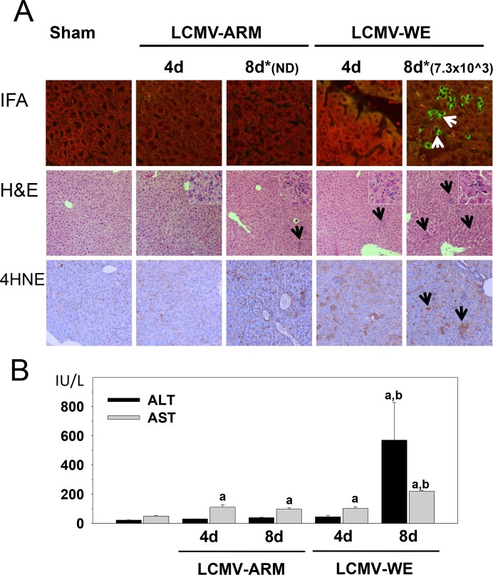
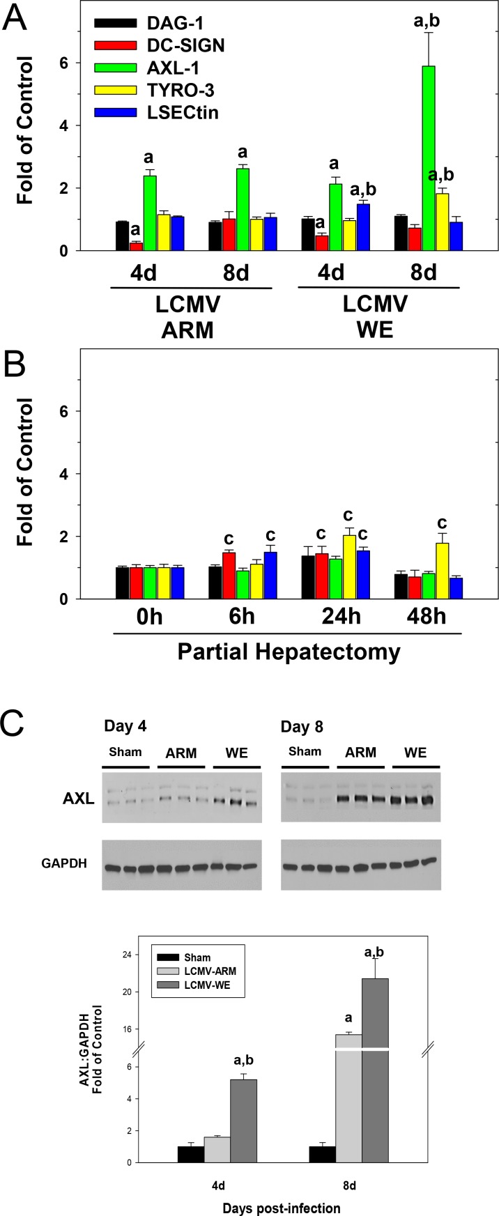
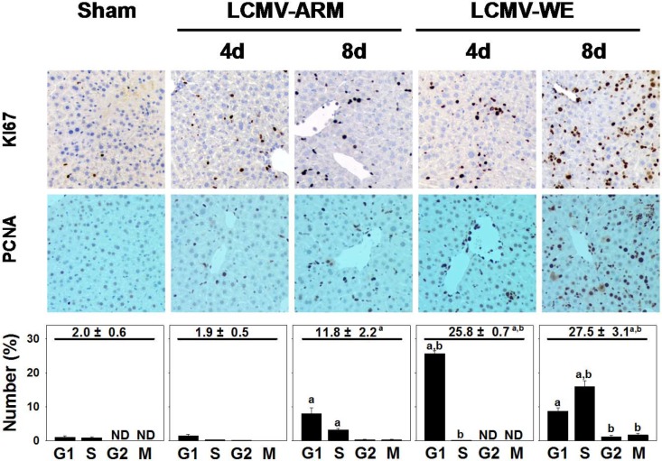
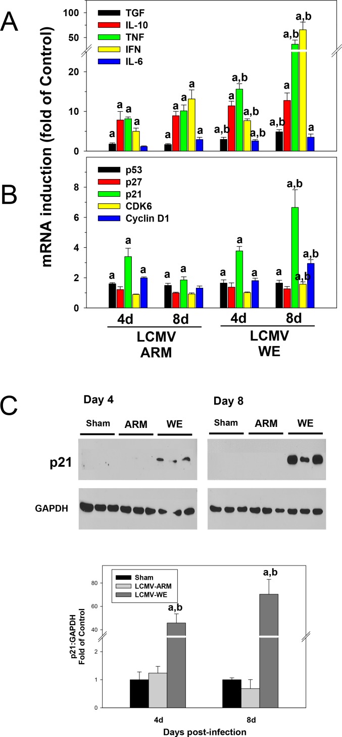
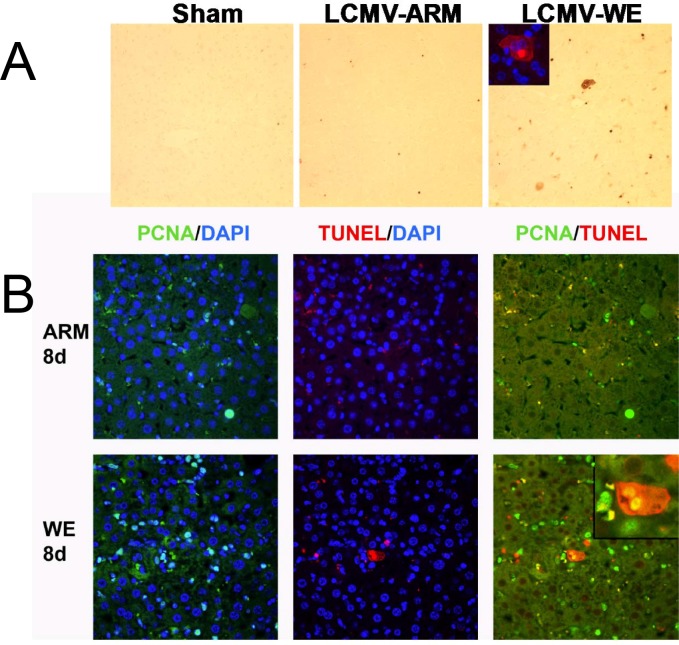
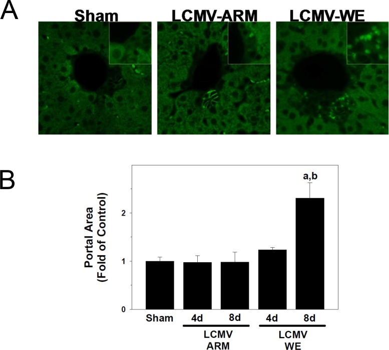
Viral hemorrhagic fevers (VHFs) encompass a group of diseases with cardinal symptoms of fever, hemorrhage, and shock. The liver is a critical mediator of VHF disease pathogenesis and high levels of ALT/AST transaminases in plasma correlate with poor prognosis. In fact, Lassa Fever (LF), the most prevalent VHF in Africa, was initially clinically described as hepatitis. Previous studies in non-human primate (NHP) models also correlated LF pathogenesis with a robust proliferative response in the liver. The purpose of the current study was to gain insight into the mechanism of liver injury and to determine the potential role of proliferation in LF pathogenesis. C57Bl/6J mice were infected with either the pathogenic (for NHPs) strain of lymphocytic choriomeningitis virus (LCMV, the prototypic arenavirus), LCMV-WE, or with the non-pathogenic strain, LCMV-ARM. As expected, LCMV-WE, but not ARM, caused a hepatitis-like infection. LCMV-WE also induced a robust increase in the number of actively cycling hepatocytes. Despite this increase in proliferation, there was no significant difference in liver size between LCMV-WE and LCMV-ARM, suggesting that cell cycle was incomplete. Indeed, cells appeared arrested in the G1 phase and LCMV-WE infection increased the number of hepatocytes that were simultaneously stained for proliferation and apoptosis. LCMV-WE infection also induced expression of a non-conventional virus receptor, AXL-1, from the TAM (TYRO3/AXL/MERTK) family of receptor tyrosine kinases and this expression correlated with proliferation. Taken together, these results shed new light on the mechanism of liver involvement in VHF pathogenesis. Specifically, it is hypothesized that the induction of hepatocyte proliferation contributes to expansion of the infection to parenchymal cells. Elevated levels of plasma transaminases are likely explained, at least in part, by abortive cell cycle arrest induced by the infection. These results may lead to the development of new therapies to prevent VHF progression.


Conflict of interest statement

Competing Interests: The authors have declared that no competing interests exist.

Figures

Fig 1
Fig 1. Transient hepatitis in LCMV-WE- infected C57BL/6J mice.
Female C57BL/6J mice were divided into two groups and intravenously infected with either LCMV-WE or with LCMV-ARM, 1×10⁶ PFU in 0.3 ml of PBS. Panel A. IFA, staining for LCMV antigen (400x magnification); H&E, staining for hematoxylin and eosin (200x); 4HNE, IHC with a monoclonal anti-4HNE IgG antibody as a marker of oxidative damage and lipid peroxidation (200x). Arrows point to positive staining (IFA and 4HNE) or inflammatory foci (H&E). Insets (1000x magnification) depict inflammatory cell recruitment in LCMV-infected livers. *, numbers above IFA staining panels are viral titer values for LCMV-WE infected liver extracts in PFU/g; ND, not determined, lower than detection limit (<1.2 log10 PFU/g, see details in Results). Panel B. Plasma alanine aminotransferase (ALT) and aspartate aminotransferase (AST) were determined as described in Materials and Methods. Data are means ± SEM (n = 4–7).a, P<0.05 compared with sham infection;b, P<0.05 compared with LCMV-ARM infection.
Fig 2
Fig 2. LCMV-WE infection upregulates expression of genes encoding non-conventional receptors for virus entry.
Hepatic mRNA was extracted from LCMV-infected mice (Panel A) or from mice after partial hepatectomy (Panel B) and real-time RT-PCR was performed as described in Materials and Methods. Western blot analysis of AXL protein expression (Panel C) was determined as described in Materials and Methods. GAPDH was used as a loading control. Representative bands (upper) and quantitative densitometry (lower) are shown. Quantitative data are means ± SEM (n = 4–7).a, P<0.05 compared with sham infection;b, P<0.05 compared with LCMV-ARM infection;c, P<0.05 compared sham surgery (t = 0; Panel B). Data are representative of two experiments performed with liver extracts collected from three mice in each group.
Fig 3
Fig 3. LCMV-WE infection causes cell cycle abortion in hepatocytes.
Immunohistochemistry for Ki-67 (200x), proliferating-cell-nuclear-antigen (PCNA, 200x) and quantitative analysis were performed as described in Materials and Methods. Quantitative PCNA summarizes the total % of cells in interphase (upper value), as well as at the individual cell cycle stages (i.e., G1, S, G2, M). Quantitative data are means ± SEM (n = 4–7).a, P<0.05 compared with sham infection;b, P<0.05 compared with LCMV-ARM infection. Data are representative of two experiments performed with liver sections from three mice in each group.
Fig 4
Fig 4. Proinflammatory cytokines and cell cycle gene responses in liver of LCMV-infected mice.
Hepatic mRNA was extracted from LCMV-infected mice and real-time RT-PCR for inflammatory (Panel A) and cell cycle (Panel B) genes was performed as described in Materials and Methods. Western blot analysis of p21 protein expression (Panel C) was determined as described in Materials and Methods. GAPDH was used as a loading control. Representative bands (upper) and quantitative densitometry (lower) are shown. Quantitative data are means ± SEM (n = 4–7).a, P<0.05 compared with sham infection;b, P<0.05 compared with LCMV-ARM infection. Data are representative of two experiments performed with liver extracts collected from three mice in each group.
Fig 5
Fig 5. Proliferation and apoptosis overlaps in livers from LCMV-WE-infected mice.
Animal groups are as described in Fig 1. Immunohistochemical detection of apoptosis (TUNEL) and proliferation (PCNA) staining were performed as described in Materials and Methods. 4',6-diamidino-2-phenylindole (DAPI, blue) nuclear staining was used as a counterstain for immunofluorescent techniques. Panel A shows representative photomicrographs (200x) depicting apoptosis; the inset (400x) depicts immunofluorescent staining for non-traditional cytosolic TUNEL staining in livers from LCMV-WE infected mice. Panel B shows representative photomicrographs (200x) depicting double immunofluorescent staining for PCNA (left; green) and TUNEL (middle; red) and merged (right; yellow) in livers from LCMV-infected mice. The inset depicts double-immunofluorescent staining in a cell with non-traditional cytosolic TUNEL staining in livers from LCMV-WE infected mice (see also inset for Panel A).
Fig 6
Fig 6. LCMV-WE causes expansion of progenitor cells in portal regions.
Animals and treatments are as described in Fig 1 and Materials and Methods. Immunofluorescent detection of A6-positive progenitor cells was performed as described in Materials and Methods. The estimation of the cellular area of the portal tract was determined by image-analysis as described in Materials and Methods and reported as fold of control (sham). Panel A shows representative photomicrographs (400x) of A6 staining in livers from sham or LCMV-infected mice. Insets show a blow-up of the A6-positive cells (or the equivalent area). Panel B shows quantiative determination of portal area size. Quantitative data are means ± SEM (n = 4–7).a, P<0.05 compared with sham infection;b, P<0.05 compared with LCMV-ARM infection.

References

    1. Crespo G, Marino Z, Navasa M, Forns X (2012) Viral Hepatitis in liver transplantation. Gastroenterology 142: 1373–1383. doi: 10.1053/j.gastro.2012.02.011 - DOI - PubMed
    1. Zhou X, Ramachandran S, Mann M, Popkin DL (2012) Role of Lymphocytic Choriomeningitis Virus (LCMV) in Understanding Viral Immunology: Past, Present and Future. Viruses 4: 2650–2669. doi: 10.3390/v4112650 - DOI - PMC - PubMed
    1. Zinkernagel R (2002) Lymphocytic choriomeningitis virus and immunology. Curr Top Microbiol Immunol 262: 1–5. - PubMed
    1. Buchmeier MJ, Welsh RM, Dutko FJ, Oldstone MBA (1980) The virology and immunobiology of lymphocytic choriomeningitis virus infection. Adv Immunol 30: 275–331. - PubMed
    1. Salvato MS, Clegg JCS, Buchmeier MJ, Charrel RN, Gonzalez JP, et al. (2012) Family Arenaviridae In: King A.M.Q., Adams M.J., Carstens E.B., Lefkowitz E.J. (Eds.), Virus Taxonomy, IXth Report of the ICTV. Elsevier/Academic Press, London: 715–723.

Publication types