. 2015 Mar 30;10(3):e0123960.
doi: 10.1371/journal.pone.0123960. eCollection 2015.

Polysialylation of NCAM characterizes the proliferation period of contractile elements during postnatal development of the epididymis

Affiliations

Polysialylation of NCAM characterizes the proliferation period of contractile elements during postnatal development of the epididymis

Peter Simon et al. PLoS One. .

Abstract

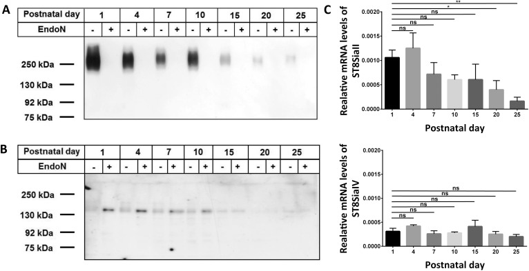
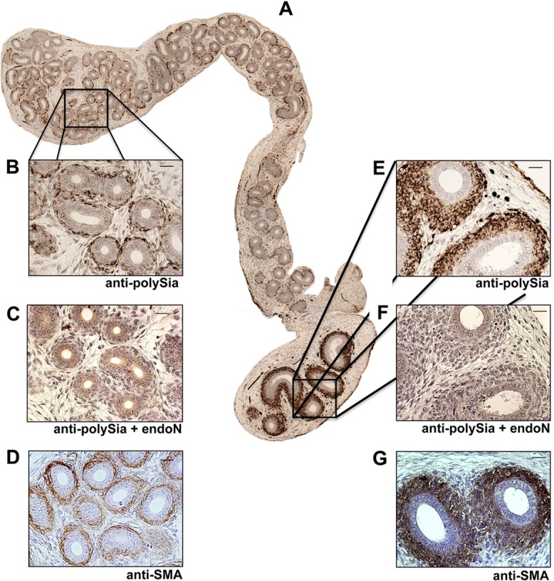
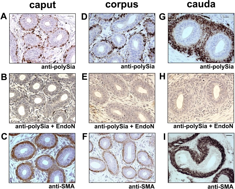
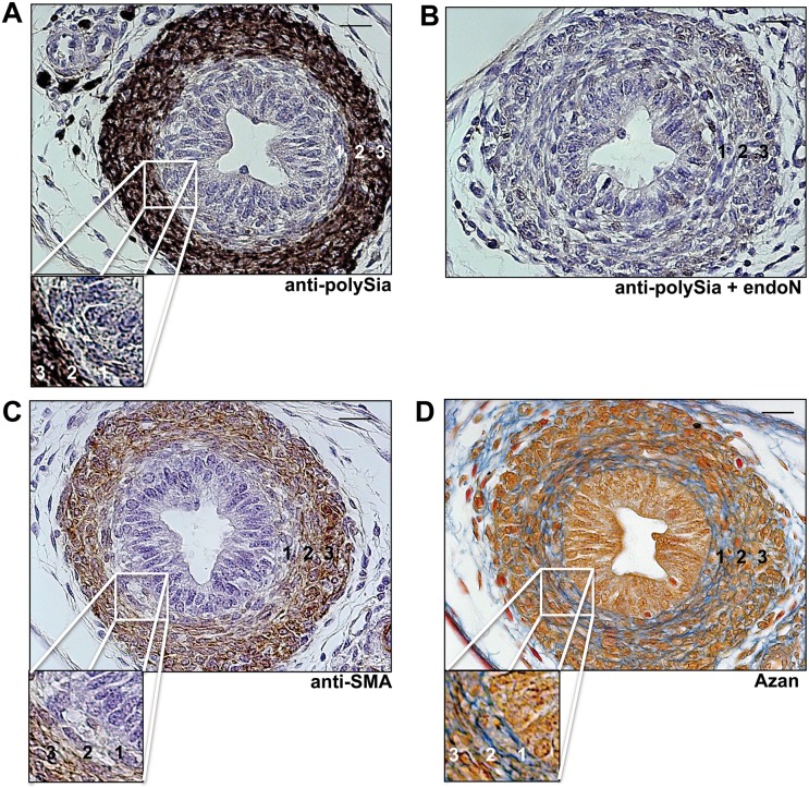
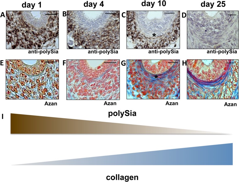
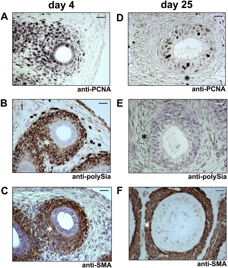
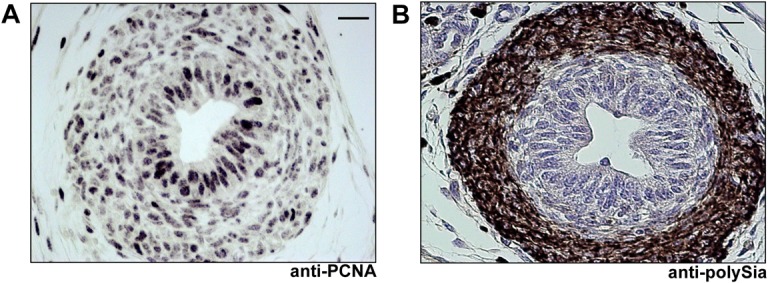
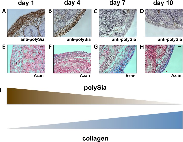
Polysialic acid (polySia) attached to the neural cell adhesion molecule (NCAM) regulates inter alia the proliferation and differentiation via the interactions with neurotrophins. Since in postnatal epididymis neurotrophins and their receptors like the Low-Affinity Nerve Growth Factor Receptor p75 and TrK B receptor are expressed, we wanted to analyze if the polysialylation of NCAM is also involved during the development of the epididymis. To this end, we monitored the developmental changes in the expression of the polysialyltransferases and NCAM polysialylation using murine epididymis at different time points during postnatal development. Our results revealed that during postnatal development of the epididymis both polysialyltransferases, ST8SiaII and ST8SiaIV, were expressed and that the expression levels dropped with increasing age. In agreement with the expression levels of the polysialyltransferases the highest content of polysialylated NCAM was present during the first 10 days after birth. Interestingly, proliferating smooth muscle cell populations prevalently expressed polysialylated NCAM. Furthermore, we observed that inverse to the decrease in polysialylation of smooth muscle cells a strong up-regulation of collagen takes place suggesting a functional relationship since collagen was recently described to induce the turnover of polysialylated NCAM via an induction of endocytosis in cellulo. The same time course of polySia and collagen synthesis was also observed in other regions of the male reproductive system e.g. vas deferens and tunica albuginea (testis). Together, we identified a spatio-temporal expression pattern of polySia-NCAM characterized by high proliferation rate of smooth muscle cells and low collagen content.


Conflict of interest statement

Competing Interests: The authors have declared that no competing interests exist.

Figures

Fig 1
Fig 1. Polysialylation status and expression levels of ST8SiaII and ST8SiaIV during postnatal development of epididymis.
Postnatal epididymis homogenates were separated by SDS-PAGE for Western blotting using 0.3 μg protein per lane with or without endoN pretreatment. Immunostaining was performed with an (A) anti-polySia mAb and (B) anti-NCAM mAb H28. The expected molecular weight of the polysialylated form of NCAM ranged between 120 and 300 kDa. Apparent molecular weights of standard proteins are indicated in kDa. (C) mRNA expression levels of ST8SiaII and ST8SiaIV in postnatal epididymis were determined by quantitative real time PCR. β-Actin was used as standard housekeeping gene to calculate the relative mRNA values of ST8SiaII and ST8SiaIV (mRNA β-Actin / mRNA polysialyltransferases). Values represent means ± SEM (standard error of the mean) of 3 independent experiments. *, P<0.05; **, P<0.01; ns = non-significant).
Fig 2
Fig 2. Immunohistological localization of polySia in epididymal tissue of 4-day-old mice.
(A-G) Paraffin-embedded serial epididymis sections. Whole longitudinal epididymis of mice were stained with a mAb against polySia (A,B,E,). For negative control, tissue sections were pretreated with endoN to degrade polySia (C,G). For identification of smooth muscle cells mAB against smooth muscle actin (D,G) were used. Tissues were counterstained with Haemalaun. Scale bars representing 20 μm.
Fig 3
Fig 3. Immunohistological localization of polySia in epididymal tissue of 10-day-old mice.
(A)-(F) Paraffin-embedded serial sections of caput, corpus and cauda epididymis. PolySia+ cells were identified with a mAb against polySia (A,D,G). For negative control, tissue sections were pretreated with endoN to degrade polySia (B,E,H). For identification of smooth muscle cells a mAb against smooth muscle actin (C,F,I) were used. Sections were counterstained with Haemalaun. Scale bars representing 20 μm.
Fig 4
Fig 4. Immunohistological localization of polySia in vas deferens of postnatal mice.
Transversal paraffin-embedded serial sections of vas deferens of 7 day old mice were stained with a mAb against polySia (A). For negative control, tissue sections were pretreated with endoN to degrade polySia (B). For identification of smooth muscle cells mAB against smooth muscle actin (C) were used as well as Azan staining (D) was performed. 1 = inner layer, 2 = middle layer, 3 = outer layer of smooth muscle cells surrounding the vas deferens. Cutouts show a higher magnification of the inner longitudinal layer of the muscle cells surrounding the vas deferens. Tissues (A-C) were counterstained with Haemalaun. Scale bars representing 20 μm.
Fig 5
Fig 5. Immunohistological analysis of polySia+cells and collagen status of the inner longitudinal layer of smooth muscle cells in early and late postnatal mice epididymis.
Paraffin-embedded serial section of epididymis (cauda) at different time points (A-H). Sections of epididymis were stained with a mAb against polySia (A-D). For negative control, tissue sections were pretreated with endoN to degrade polySia (data not shown). An anti-SMA immunostaining was used to identify smooth muscle cells (data not shown). For collagen identification Azan staining was performed (E-H). * labels same positions of smooth muscle cells. Tissues were counterstained with Haemalaun (A-D). Scale bars representing 20 μm. (I) Illustration of the inverse correlation between polySia and collagen.
Fig 6
Fig 6. Immunohistological analysis of polySia+cells and cell proliferation status in early and late postnatal mice epididymis.
Paraffin-embedded serial section of 4 days old (A-C) and 25 days old (D-F) mouse epididymis (cauda). Sections were stained with a mAb against proliferating cell nuclear antigen (anti-PCNA) for proliferating status (A,D) and additional stained with a mAb against polySia (B,E). For negative control, tissue sections were pretreated with endoN to degrade polySia (data not shown). For identification of smooth muscle cells a mAB against smooth muscle actin were used (C,F). * labels same positions of smooth muscle cells. Tissues were counterstained with Haemalaun. Scale bars representing 20 μm.
Fig 7
Fig 7. Immunohistological analysis of polySia+cells and cell proliferation status in postnatal vas deferens.
Paraffin-embedded serial sections of 7 days old vas deferens were stained with a mAb against proliferating cell nuclear antigen (anti-PCNA) (A) and a mAb against polySia (B). For negative control tissue sections were pretreated with endoN to degrade polySia (data not shown). Tissues were counterstained with Haemalaun. Scale bars representing 20 μm.
Fig 8
Fig 8. Immunohistological analysis of polySia+cells and collagen status of the tunica albuginea in early and late postnatal mice testis.
Paraffin-embedded serial section of testis were stained with a mAb against polySia (A-D). For negative control, tissue sections were pretreated with endoN to degrade polySia (data not shown). For collagen visualization Azan staining was performed (E-H). Tissues were counterstained with Haemalaun (A-D). Scale bars representing 20 μm. (I) Illustration of the inverse correlation between polySia and collagen.

References

    1. Colley KJ. Structural basis for the polysialylation of the neural cell adhesion molecule. Adv Exp Med Biol 2010;663: 111–126. 10.1007/978-1-4419-1170-4_7 - DOI - PubMed
    1. Sato C, Kitajima K. Disialic, oligosialic and polysialic acids: distribution, functions and related disease. J Biochem 2013;154: 115–136. 10.1093/jb/mvt057 - DOI - PubMed
    1. Mühlenhoff M, Rollenhagen M, Werneburg S, Gerardy-Schahn R, Hildebrandt H. Polysialic acid: versatile modification of NCAM, SynCAM 1 and neuropilin-2. Neurochem Res 2013;38: 1134–1143. 10.1007/s11064-013-0979-2 - DOI - PubMed
    1. Simon P, Bäumner S, Busch O, Röhrich R, Kaese M, Richterich P, et al. Polysialic Acid Is Present in Mammalian Semen as a Post-translational Modification of the Neural Cell Adhesion Molecule NCAM and the Polysialyltransferase ST8SiaII. J Biol Chem 2013;288: 18825–18833. 10.1074/jbc.M113.451112 - DOI - PMC - PubMed
    1. Galuska SP, Geyer R, Gerardy-Schahn R, Mühlenhoff M, Geyer H. Enzyme-dependent Variations in the Polysialylation of the Neural Cell Adhesion Molecule (NCAM) in Vivo. J Biol Chem 2008;283: 17–28. - PubMed

Publication types